Explore
 Validate
 Learn
Learn Western blot
 ELISA
ELISA Other assay
Antibody data
- Antibody Data
- Antigen structure
- References [4]
- Comments [0]
- Validations
- Other assay [2]
Submit
Validation data
Reference
Comment
Report error
- Product number
- PA1-931 - Provider product page

- Provider
- Invitrogen Antibodies
- Product name
- Calbindin D28K Polyclonal Antibody
- Antibody type
- Polyclonal
- Antigen
- Purifed from natural sources
- Description
- PA1-931 detects calbindin D-28k protein in rat samples. PA1-931 has successfully been used in Western blot , ELISA, immunoprecipitation and immunohistochemical procedures. By Western blot, this antibody detects a 28 kDa protein representing calbindin D from rat cerebellum and rat kidney extract. Immunohistochemical staining on paraffin sections demonstrates that calbindin D is localized to the cytoplasm in epithelial cells of distal tubules in human kidney cortex after staining with PA1-931. The PA1-931 immunizing protein corresponds to the purified 28 kDa calbindin-D protein from rat kidney. Reconstitute in 100 µL PBS to create a stock of 1 mg/mL.
- Reactivity
- Human, Mouse, Rat
- Host
- Rabbit
- Isotype
- IgG
- Vial size
- 100 μg
- Concentration
- 1 mg/mL
- Storage
- -20°C, Avoid Freeze/Thaw Cycles
Submitted references Conditional Deletion of Activating Rearranged During Transfection Receptor Tyrosine Kinase Leads to Impairment of Photoreceptor Ribbon Synapses and Disrupted Visual Function in Mice.
Lycii radicis cortex inhibits glucocorticoid‑induced bone loss by downregulating Runx2 and BMP‑2 expression.
Anti-Yo antibody uptake and interaction with its intracellular target antigen causes Purkinje cell death in rat cerebellar slice cultures: a possible mechanism for paraneoplastic cerebellar degeneration in humans with gynecological or breast cancers.
Developmental and age-dependent changes of 28-kDa calbindin-D in the central nervous tissue determined with a sensitive immunoassay method.
Peng WH, Liao ML, Huang WC, Liu PK, Levi SR, Tseng YJ, Lee CY, Yeh LK, Chen KJ, Chien CL, Wang NK
Frontiers in neuroscience 2021;15:728905
Frontiers in neuroscience 2021;15:728905
Lycii radicis cortex inhibits glucocorticoid‑induced bone loss by downregulating Runx2 and BMP‑2 expression.
Lee B, Hong S, Kim M, Kim EY, Park HJ, Jung HS, Kim JH, Sohn Y
International journal of molecular medicine 2021 Aug;48(2)
International journal of molecular medicine 2021 Aug;48(2)
Anti-Yo antibody uptake and interaction with its intracellular target antigen causes Purkinje cell death in rat cerebellar slice cultures: a possible mechanism for paraneoplastic cerebellar degeneration in humans with gynecological or breast cancers.
Greenlee JE, Clawson SA, Hill KE, Wood B, Clardy SL, Tsunoda I, Carlson NG
PloS one 2015;10(4):e0123446
PloS one 2015;10(4):e0123446
Developmental and age-dependent changes of 28-kDa calbindin-D in the central nervous tissue determined with a sensitive immunoassay method.
Kurobe N, Inaguma Y, Shinohara H, Semba R, Inagaki T, Kato K
Journal of neurochemistry 1992 Jan;58(1):128-34
Journal of neurochemistry 1992 Jan;58(1):128-34
No comments: Submit comment
Supportive validation
- Submitted by
- Invitrogen Antibodies (provider)
- Main image

- Experimental details
- NULL
- Submitted by
- Invitrogen Antibodies (provider)
- Main image
- Experimental details
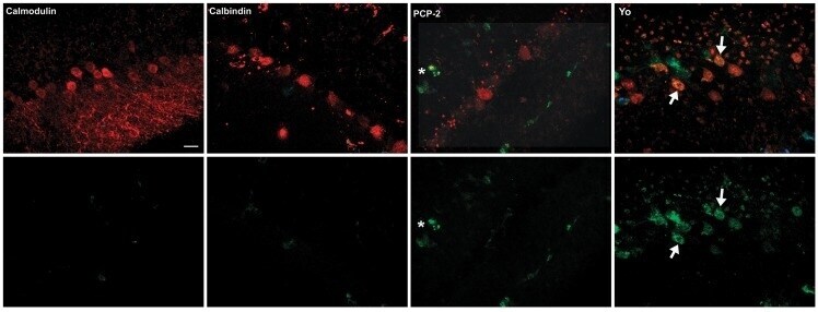
- Fig 4 Comparison of uptake and cytotoxicity of anti-Yo antibody versus three other antibodies specific for the intracellular Purkinje cell proteins: calbindin, calmodulin, and PCP-2. The top row of panels demonstrates uptake and accumulation of IgG (red) within Purkinje cells after 96 hours in cultures incubated with anti-calmodulin, anti-calbindin, anti PCP-2, or anti-Yo IgGs. Entry of SYTOX green into Purkinje cells containing IgG, indicative of cell membrane injury and death (yellow), was seen in only in cultures incubated with anti-Yo IgGs (examples shown by arrows). The lower panels show only SYTOX green staining of Purkinje cells in cultures incubated with the antibodies indicated. In the culture incubated with anti-PCP-2. SYTOX staining indicative of cell death is seen in a single cell outside the Purkinje cell layer (asterisk) but not in Purkinje cells. Cultures were followed through 144 hours. There was progression of cell death in cultures incubated with anti-Yo antibodies. In contrast, cultures incubated calbindin, calmodulin, and PCP-2 did not exhibit Purkinje cell death above background levels seen in controls incubated with normal human IgG (data not shown). Scale bar = 20mu.