Explore
 Validate
 Learn
LearnMA1-22165
antibody from Invitrogen Antibodies
Targeting: SELE
CD62E, ELAM, ELAM1, ESEL
 Western blot ELISA
 Immunocytochemistry Immunoprecipitation Immunohistochemistry Flow cytometry Other assay
Antibody data
- Antibody Data
- Antigen structure
- References [4]
- Comments [0]
- Validations
- Immunocytochemistry [2]
- Flow cytometry [2]
- Other assay [3]
Submit
Validation data
Reference
Comment
Report error
- Product number
- MA1-22165 - Provider product page

- Provider
- Invitrogen Antibodies
- Product name
- E-selectin Monoclonal Antibody (1.2B6)
- Antibody type
- Monoclonal
- Antigen
- Other
- Description
- Not suitable for immunohistochemistry (paraffin). Store product as a concentrated solution. Centrifuge briefly prior to opening the vial.
- Reactivity
- Human, Porcine
- Host
- Mouse
- Isotype
- IgG
- Antibody clone number
- 1.2B6
- Vial size
- 100 μg
- Concentration
- 1 mg/mL
- Storage
- Store at 4°C short term. For long term storage, store at -20°C, avoiding freeze/thaw cycles.
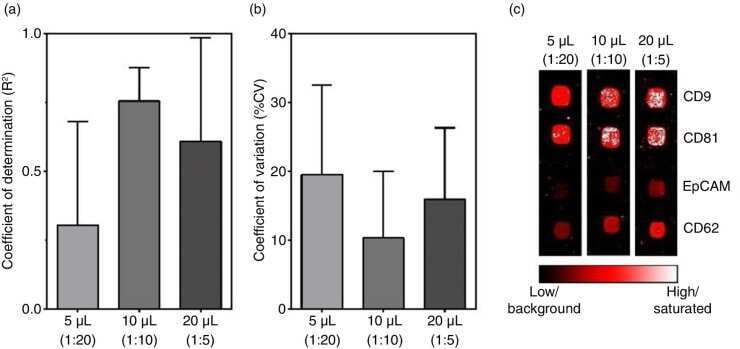
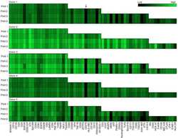
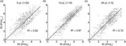
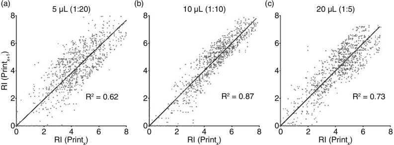
Submitted references The impact of various preanalytical treatments on the phenotype of small extracellular vesicles in blood analyzed by protein microarray.
Potentials and capabilities of the Extracellular Vesicle (EV) Array.
Potentials and capabilities of the Extracellular Vesicle (EV) Array.
Novel anti-inflammatory properties of the angiogenesis inhibitor vasostatin.
Bæk R, Søndergaard EK, Varming K, Jørgensen MM
Journal of immunological methods 2016 Nov;438:11-20
Journal of immunological methods 2016 Nov;438:11-20
Potentials and capabilities of the Extracellular Vesicle (EV) Array.
Jørgensen MM, Bæk R, Varming K
Journal of extracellular vesicles 2015;4:26048
Journal of extracellular vesicles 2015;4:26048
Potentials and capabilities of the Extracellular Vesicle (EV) Array.
Jørgensen MM, Bæk R, Varming K
Journal of extracellular vesicles 2015;4:26048
Journal of extracellular vesicles 2015;4:26048
Novel anti-inflammatory properties of the angiogenesis inhibitor vasostatin.
Huegel R, Velasco P, De la Luz Sierra M, Christophers E, Schröder JM, Schwarz T, Tosato G, Lange-Asschenfeldt B
The Journal of investigative dermatology 2007 Jan;127(1):65-74
The Journal of investigative dermatology 2007 Jan;127(1):65-74
No comments: Submit comment
Supportive validation
- Submitted by
- Invitrogen Antibodies (provider)
- Main image

- Experimental details
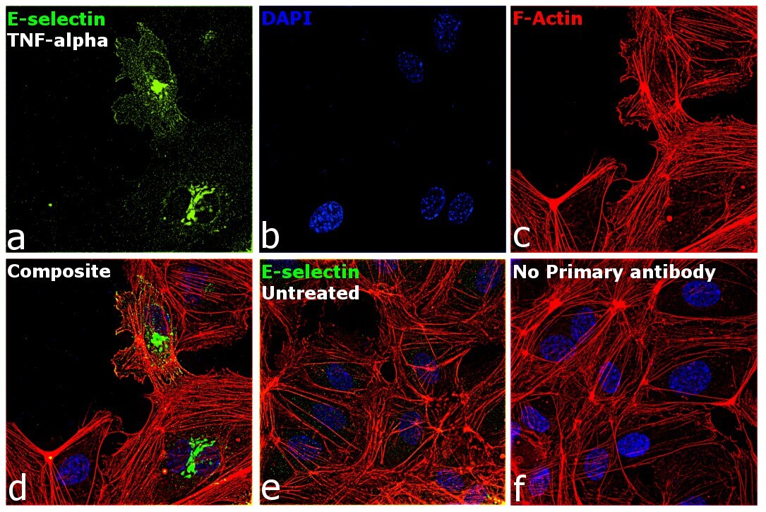
- Immunofluorescence analysis of E-selectin was performed using 70% confluent log phase HUVEC cells treated with 100 U/mL TNF-alpha for 3 Hrs. The cells were fixed with 4% Paraformaldehyde for 10 minutes, permeabilized with 0.1% Triton™ X-100 for 10 minutes, and blocked with 2% BSA for 10 minutes at room temperature. The cells were labeled with E-selectin Monoclonal Antibody (1.2B6) (Product # MA1-22165) at 1:100 dilution in 0.1% BSA, incubated at 4 degree celsius overnight and then labeled with Donkey anti-Mouse IgG (H+L) Highly Cross-Adsorbed Secondary Antibody, Alexa Fluor Plus 488 (Product # A32766, 1:2000 dilution) for 45 minutes at room temperature (Panel a: Green). Nuclei (Panel b: Blue) were stained with SlowFade® Gold Antifade Mountant with DAPI (Product # S36938). F-actin (Panel c: Red) was stained with Rhodamine Phalloidin (Product # R415, 1:300). Panel d represents the merged image showing cytoplasmic and membrane localization. Panel e represents untreated cells with reduced signal. Panel f represents control cells with no primary antibody to assess background. The images were captured at 60X magnification.
- Submitted by
- Invitrogen Antibodies (provider)
- Main image
- Experimental details
- Immunofluorescence analysis of E-selectin was performed using 70% confluent log phase HUVEC cells treated with 100 U/mL TNF-alpha for 3 Hrs. The cells were fixed with 4% Paraformaldehyde for 10 minutes, permeabilized with 0.1% Triton™ X-100 for 10 minutes, and blocked with 2% BSA for 10 minutes at room temperature. The cells were labeled with E-selectin Monoclonal Antibody (1.2B6) (Product # MA1-22165) at 1:100 dilution in 0.1% BSA, incubated at 4 degree celsius overnight and then labeled with Donkey anti-Mouse IgG (H+L) Highly Cross-Adsorbed Secondary Antibody, Alexa Fluor Plus 488 (Product # A32766, 1:2000 dilution) for 45 minutes at room temperature (Panel a: Green). Nuclei (Panel b: Blue) were stained with SlowFade® Gold Antifade Mountant with DAPI (Product # S36938). F-actin (Panel c: Red) was stained with Rhodamine Phalloidin (Product # R415, 1:300). Panel d represents the merged image showing cytoplasmic and membrane localization. Panel e represents untreated cells with reduced signal. Panel f represents control cells with no primary antibody to assess background. The images were captured at 60X magnification.

Supportive validation
- Submitted by
- Invitrogen Antibodies (provider)
- Main image

- Experimental details
- Flow Cytometry analysis of Collagen I was performed in thrombin activated human peripheral blood platelets using Collagen I Polyclonal Antibody, Biotin (Product # PA1-28530).

- Submitted by
- Invitrogen Antibodies (provider)
- Main image

- Experimental details
- Flow Cytometry analysis of Collagen I was performed in thrombin activated human peripheral blood platelets using Collagen I Polyclonal Antibody, Biotin (Product # PA1-28530).

Supportive validation
- Submitted by
- Invitrogen Antibodies (provider)
- Main image
- Experimental details
- NULL

- Submitted by
- Invitrogen Antibodies (provider)
- Main image
- Experimental details
- NULL
- Submitted by
- Invitrogen Antibodies (provider)
- Main image
- Experimental details
- NULL