Explore
 Validate
 Learn
Learn Flow cytometry
Antibody data
- Antibody Data
- Antigen structure
- References [5]
- Comments [0]
- Validations
- Flow cytometry [1]
- Other assay [1]
Submit
Validation data
Reference
Comment
Report error
- Product number
- 12-0627-41 - Provider product page

- Provider
- Invitrogen Antibodies
- Product name
- CD62E (E-selectin) Monoclonal Antibody (P2H3), PE, eBioscience™
- Antibody type
- Monoclonal
- Antigen
- Other
- Description
- Description: The P2H3 monoclonal antibody reacts with human CD62E, a 97-115 kDa member of the selectin family. CD62E, also known as E-selectin or endothelial-leukocyte adhesion molecule-1 (ELAM-1) is an adhesion molecule expressed by endothelial cells upon stimulation with cytokines including TNFalpha and IL-1beta. Induced expression of CD62E during inflammatory conditions is thought to mediate leukocyte rolling including the initial interaction of neutrophils with endothelium.
- Conjugate
- Yellow dye
- Antibody clone number
- P2H3
- Concentration
- 5 µL/Test
Submitted references Colonization of dermal arterioles by Neisseria meningitidis provides a safe haven from neutrophils.
Expansion of functional personalized cells with specific transgene combinations.
Effect of nicotine and porphyromonas gingivalis lipopolysaccharide on endothelial cells in vitro.
Lipopolysaccharide and sphingosine-1-phosphate cooperate to induce inflammatory molecules and leukocyte adhesion in endothelial cells.
Detection and monitoring of the multiple inflammatory responses by photoacoustic molecular imaging using selectively targeted gold nanorods.
Manriquez V, Nivoit P, Urbina T, Echenique-Rivera H, Melican K, Fernandez-Gerlinger MP, Flamant P, Schmitt T, Bruneval P, Obino D, Duménil G
Nature communications 2021 Jul 27;12(1):4547
Nature communications 2021 Jul 27;12(1):4547
Expansion of functional personalized cells with specific transgene combinations.
Lipps C, Klein F, Wahlicht T, Seiffert V, Butueva M, Zauers J, Truschel T, Luckner M, Köster M, MacLeod R, Pezoldt J, Hühn J, Yuan Q, Müller PP, Kempf H, Zweigerdt R, Dittrich-Breiholz O, Pufe T, Beckmann R, Drescher W, Riancho J, Sañudo C, Korff T, Opalka B, Rebmann V, Göthert JR, Alves PM, Ott M, Schucht R, Hauser H, Wirth D, May T
Nature communications 2018 Mar 8;9(1):994
Nature communications 2018 Mar 8;9(1):994
Effect of nicotine and porphyromonas gingivalis lipopolysaccharide on endothelial cells in vitro.
An N, Andrukhov O, Tang Y, Falkensammer F, Bantleon HP, Ouyang X, Rausch-Fan X
PloS one 2014;9(5):e96942
PloS one 2014;9(5):e96942
Lipopolysaccharide and sphingosine-1-phosphate cooperate to induce inflammatory molecules and leukocyte adhesion in endothelial cells.
Fernández-Pisonero I, Dueñas AI, Barreiro O, Montero O, Sánchez-Madrid F, García-Rodríguez C
Journal of immunology (Baltimore, Md. : 1950) 2012 Dec 1;189(11):5402-10
Journal of immunology (Baltimore, Md. : 1950) 2012 Dec 1;189(11):5402-10
Detection and monitoring of the multiple inflammatory responses by photoacoustic molecular imaging using selectively targeted gold nanorods.
Ha S, Carson A, Agarwal A, Kotov NA, Kim K
Biomedical optics express 2011 Feb 23;2(3):645-57
Biomedical optics express 2011 Feb 23;2(3):645-57
No comments: Submit comment
Supportive validation
- Submitted by
- Invitrogen Antibodies (provider)
- Main image

- Experimental details
- Human Umbilical Vein Endothelial Cells (HUVEC) were activated with Human TNF alpha Recombinant Protein (Product # 14-8329-81) and stained with Mouse IgG1 kappa Isotype Control PE (Product # 12-4714-81) (open histogram) or Anti-Human CD62E (E-Selectin) PE (filled histogram). Total viable cells were used for analysis.
- Conjugate
- Yellow dye

Supportive validation
- Submitted by
- Invitrogen Antibodies (provider)
- Main image

- Experimental details
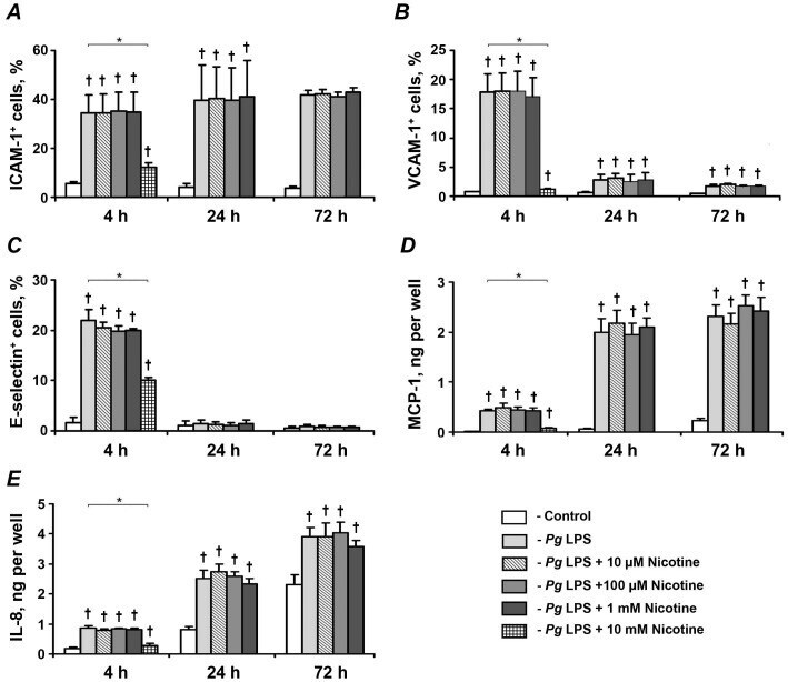
- Figure 5 Effect of nicotine on the P. gingivalis LPS-induced protein expression of pro-inflammatory mediators in HUVECs. HUVECs were stimulated by P. gingivalis LPS in the presence or absence of nicotine (10 uM-10 mM) for 4, 24, and 72 h. After stimulation, the surface expression levels of ICAM-1 (A), VCAM-1 (B), and E-selectin (C) were measured by flow cytometry, and the quantity of MCP-1 (D) and IL-8 (E) in conditioned media was measured by ELISA. Each value represents mean +-SD of three independent assays. Non-stimulated HUVECs were used as a control. The protein expression levels of pro-inflammatory mediators were not analyzed after stimulation with 10-mM nicotine for 24 and 72 h because the cells were not viable. * - significantly different between groups, p
- Conjugate
- Yellow dye