 Explore
 Validate
 Learn
Learn Western blot
Antibody data
- Antibody Data
- Antigen structure
- References [2]
- Comments [0]
- Validations
- Western blot [1]
- Immunohistochemistry [1]
- Flow cytometry [1]
Submit
Validation data
Reference
Comment
Report error
- Product number
- AF6480 - Provider product page

- Provider
- Novus Biologicals
- Product name
- Sheep Polyclonal CEACAM1/CD66a Antibody
- Antibody type
- Polyclonal
- Description
- Antigen Affinity-purified. Detects mouse CEACAM-1/CD66a in Western blots.
- Reactivity
- Mouse
- Host
- Sheep
- Conjugate
- Unconjugated
- Isotype
- IgG
- Vial size
- 100 ug
- Concentration
- LYOPH
- Storage
- Use a manual defrost freezer and avoid repeated freeze-thaw cycles. 12 months from date of receipt, -20 to -70 degreesC as supplied. 1 month, 2 to 8 degreesC under sterile conditions after reconstitution. 6 months, -20 to -70 degreesC under sterile conditions after reconstitution.
Submitted references Development of A New Mouse Model for Intrahepatic Cholangiocellular Carcinoma: Accelerating Functions of Pecam-1.
One Year Follow-Up Risk Assessment in SKH-1 Mice and Wounds Treated with an Argon Plasma Jet.
Malik IA, Malik G, Ströbel P, Wilting J
Cancers 2019 Jul 24;11(8)
Cancers 2019 Jul 24;11(8)
One Year Follow-Up Risk Assessment in SKH-1 Mice and Wounds Treated with an Argon Plasma Jet.
Schmidt A, Woedtke TV, Stenzel J, Lindner T, Polei S, Vollmar B, Bekeschus S
International journal of molecular sciences 2017 Apr 19;18(4)
International journal of molecular sciences 2017 Apr 19;18(4)
No comments: Submit comment
Supportive validation
- Submitted by
- Novus Biologicals (provider)
- Main image
- Experimental details
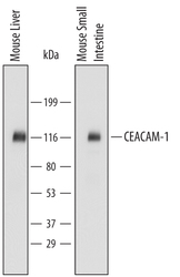
- Detection of Mouse CEACAM-1/CD66a by Western Blot. Western blot shows lysates of mouse liver tissue and mouse small intestine tissue. PVDF Membrane was probed with 0.1 µg/mL of Mouse CEACAM-1/CD66a Antigen Affinity-purified Polyclonal Antibody (Catalog # AF6480) followed by HRP-conjugated Anti-Sheep IgG Secondary Antibody (Catalog # HAF016). Specific bands were detected for CEACAM-1/CD66a between approximately 110 and 120 kDa (as indicated). This experiment was conducted under reducing conditions and using Immunoblot Buffer Group 1.

Supportive validation
- Submitted by
- Novus Biologicals (provider)
- Main image
- Experimental details
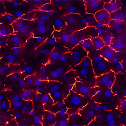
- CEACAM-1/CD66a in Mouse Liver. CEACAM-1/CD66a was detected in perfusion fixed frozen sections of mouse liver using Sheep Anti-Mouse CEACAM-1/CD66a Antigen Affinity-purified Polyclonal Antibody (Catalog # AF6480) at 5 µg/mL overnight at 4 °C. Tissue was stained using the NorthernLights™ 557-conjugated Anti-Sheep IgG Secondary Antibody (red; Catalog # NL010) and counterstained with DAPI (blue). Specific staining was localized to endothelial cells in bile canaliculi. View our protocol for Fluorescent IHC Staining of Frozen Tissue Sections.

Supportive validation
- Submitted by
- Novus Biologicals (provider)
- Main image

- Experimental details
- Detection of CEACAM-1 in Mouse Splenocytes by Flow Cytometry. Mouse splenocytes were stained with Sheep Anti-Mouse CEACAM-1/CD66a Antigen Affinity-purified Polyclonal Antibody (Catalog # AF6480) followed by Phycoerythrin-conjugated Anti-Sheep IgG Secondary Antibody (Catalog # F0126) and Rat Anti-Mouse B220/CD45R APC-conjugated Monoclonal Antibody (Catalog # FAB1217A). Quadrant markers were set based on control antibody staining (Catalog # 5-001-A).
