Explore
 Validate
 Learn
Learn Western blot
 ELISA
ELISAAntibody data
- Antibody Data
- Antigen structure
- References [2]
- Comments [0]
- Validations
- Western blot [1]
Submit
Validation data
Reference
Comment
Report error
- Product number
- A01010-2 - Provider product page

- Provider
- Boster Biological Technology
- Product name
- Anti-Complement C9 Antibody Picoband™
- Antibody type
- Polyclonal
- Description
- Polyclonal antibody for Complement Component C9/C9 detection. Host: Rabbit.Size: 100μg/vial. Tested applications: ELISA. Reactive species: Human. Complement Component C9/C9 information: Molecular Weight: 63173 MW; Subcellular Localization: Secreted. Cell membrane; Multi-pass membrane protein. Secreted as soluble monomer. Oligomerizes at target membranes, forming a pre-pore. A conformation change then leads to the formation of a 100 Angstrom diameter pore; Tissue Specificity: Plasma.
- Reactivity
- Human, Mouse
- Host
- Rabbit
- Vial size
- 100μg/vial
- Concentration
- 0.5-1mg/ml, actual concentration vary by lot. Use suggested dilution ratio to decide dilution procedure.
- Storage
- At -20°C for one year. After reconstitution, at 4°C for one month. It can also be aliquoted and stored frozen at -20°C for a longer time. Avoid repeated freezing and thawing.
- Handling
- Add 0.2ml of distilled water will yield a concentration of 500ug/ml.
Submitted references Complement factor B knockdown by short hairpin RNA inhibits laser-induced choroidal neovascularization in rats.
Evidence of extensive atherosclerosis, coronary artery disease and myocardial infarction in the ApoE(-/-):Ins2(+/Akita) mouse fed a western diet.
Wang X, Shang QL, Ma JX, Liu SX, Wang CX, Ma C
International journal of ophthalmology 2020;13(3):382-389
International journal of ophthalmology 2020;13(3):382-389
Evidence of extensive atherosclerosis, coronary artery disease and myocardial infarction in the ApoE(-/-):Ins2(+/Akita) mouse fed a western diet.
Venegas-Pino DE, Lagrotteria A, Wang PW, Morphet J, Clapdorp C, Shi Y, Werstuck GH
Atherosclerosis 2018 Aug;275:88-96
Atherosclerosis 2018 Aug;275:88-96
No comments: Submit comment
Supportive validation
- Submitted by
- Boster Biological Technology (provider)
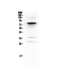
- Main image
- Experimental details
- Western blot analysis of Complement C9 using anti-Complement C9 antibody (A01010-2). Electrophoresis was performed on a 5-20% SDS-PAGE gel at 70V (Stacking gel) / 90V (Resolving gel) for 2-3 hours. The sample well of each lane was loaded with 50ug of sample under reducing conditions. Lane 1: mouse liver tissue lysates, Lane 2: mouse lung tissue lysates, After Electrophoresis, proteins were transferred to a Nitrocellulose membrane at 150mA for 50-90 minutes. Blocked the membrane with 5% Non-fat Milk/ TBS for 1.5 hour at RT. The membrane was incubated with rabbit anti-Complement C9 antigen affinity purified polyclonal antibody (Catalog # A01010-2) at 0.5 μg/mL overnight at 4°C, then washed with TBS-0.1%Tween 3 times with 5 minutes each and probed with a goat anti-rabbit IgG-HRP secondary antibody at a dilution of 1:10000 for 1.5 hour at RT. The signal is developed using an Enhanced Chemiluminescent detection (ECL) kit (Catalog # EK1002) with Tanon 5200 system. A specific band was detected for Complement C9 at approximately 80KD. The expected band size for Complement C9 is at 63KD.
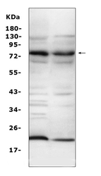
- Additional image