Explore
 Validate
 Learn
Learn Western blot
 Immunocytochemistry
Antibody data
- Antibody Data
- Antigen structure
- References [2]
- Comments [0]
- Validations
- Immunocytochemistry [2]
Submit
Validation data
Reference
Comment
Report error
- Product number
- PA5-29093 - Provider product page

- Provider
- Invitrogen Antibodies
- Product name
- Complement C9 Polyclonal Antibody
- Antibody type
- Polyclonal
- Antigen
- Recombinant full-length protein
- Description
- Recommended positive controls: THP-1, BCL-1, human plasma. Store product as a concentrated solution. Centrifuge briefly prior to opening the vial.
- Reactivity
- Human, Mouse, Rat
- Host
- Rabbit
- Isotype
- IgG
- Vial size
- 100 μL
- Concentration
- 0.62 mg/mL
- Storage
- Store at 4°C short term. For long term storage, store at -20°C, avoiding freeze/thaw cycles.
Submitted references GDF15 Suppresses Lymphoproliferation and Humoral Autoimmunity in a Murine Model of Systemic Lupus Erythematosus.
Proteomic analysis of human synovial fluid reveals potential diagnostic biomarkers for ankylosing spondylitis.
Lorenz G, Ribeiro A, von Rauchhaupt E, Würf V, Schmaderer C, Cohen CD, Vohra T, Anders HJ, Lindenmeyer M, Lech M
Journal of innate immunity 2022;14(6):673-689
Journal of innate immunity 2022;14(6):673-689
Proteomic analysis of human synovial fluid reveals potential diagnostic biomarkers for ankylosing spondylitis.
Lee JH, Jung JH, Kim J, Baek WK, Rhee J, Kim TH, Kim SH, Kim KP, Son CN, Kim JS
Clinical proteomics 2020;17:20
Clinical proteomics 2020;17:20
No comments: Submit comment
Supportive validation
- Submitted by
- Invitrogen Antibodies (provider)
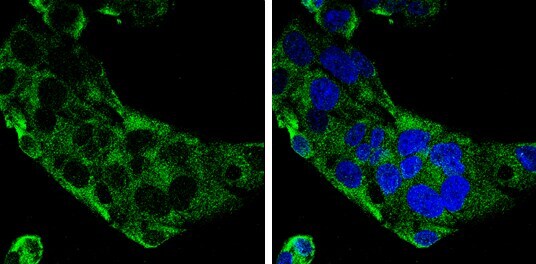
- Main image

- Experimental details
- Immunocytochemistry-Immunofluorescence analysis of Complement C9 was performed in HepG2 cells fixed in 4% paraformaldehyde at RT for 15 min. Green: Complement C9 Polyclonal Antibody (Product # PA5 29093) diluted at 1:500. Blue: Hoechst 33342 staining.

- Submitted by
- Invitrogen Antibodies (provider)
- Main image
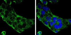
- Experimental details
- Immunocytochemistry-Immunofluorescence analysis of Complement C9 was performed in HepG2 cells fixed in 4% paraformaldehyde at RT for 15 min. Green: Complement C9 Polyclonal Antibody (Product # PA5 29093) diluted at 1:500. Blue: Hoechst 33342 staining.