Explore
 Validate
 Learn
Learn Western blot
 Immunohistochemistry
Antibody data
- Antibody Data
- Antigen structure
- References [21]
- Comments [0]
- Validations
- Immunohistochemistry [2]
- Flow cytometry [6]
Submit
Validation data
Reference
Comment
Report error
- Product number
- NBP2-29429 - Provider product page

- Provider
- Novus Biologicals
- Product name
- Mouse Monoclonal Cytokeratin, pan Antibody
- Antibody type
- Monoclonal
- Description
- Protein A or G purified. Twenty human keratins are resolved with two-dimensional gel electrophoresis into acidic (pI 6.0) subfamilies. This antibody cocktail recognizes acidic (Type I or LMW) and basic (Type II or HMW) cytokeratins, which include CK1, CK3, CK4, CK5, CK6, CK8, CK10, CK14, CK15, CK16, and CK19. Many studies have shown the usefulness of keratins as markers in cancer research and tumor diagnosis. AE-1/AE-3 is a broad spectrum anti pan-cytokeratin antibody cocktail, which differentiates epithelial tumors from non-epithelial tumors e.g. squamous vs. adenocarcinoma of the lung, liver carcinoma, breast cancer, and esophageal cancer. It has been used to characterize the source of various neoplasms and to study the distribution of cytokeratin containing cells in epithelia during normal development and during the development of epithelial neoplasms. This antibody stains cytokeratins present in normal and abnormal human tissues and has shown high sensitivity in the recognition of epithelial cells and carcinomas.
- Reactivity
- Human, Mouse, Rat, Bovine, Canine, Chicken/Avian, Rabbit, Simian, Zebrafish
- Host
- Mouse
- Isotype
- IgG
- Vial size
- 0.1 mg
- Concentration
- 0.2 mg/ml
- Storage
- Store at 4C.
Submitted references Dysregulated Expression of the Nuclear Exosome Targeting Complex Component Rbm7 in Nonhematopoietic Cells Licenses the Development of Fibrosis.
A novel immunocompetent model of metastatic prostate cancer-induced bone pain.
Adhesion G-protein-coupled receptor, GPR56, is required for Müllerian duct development in the chick.
Oncogenic KRAS-Driven Metabolic Reprogramming in Pancreatic Cancer Cells Utilizes Cytokines from the Tumor Microenvironment.
Insights into Gonadal Sex Differentiation Provided by Single-Cell Transcriptomics in the Chicken Embryo.
Polyurethane scaffolds seeded with autologous cells can regenerate long esophageal gaps: An esophageal atresia treatment model.
Cancer immunophenotyping by seven-colour multispectral imaging without tyramide signal amplification.
Loss of atrx cooperates with p53-deficiency to promote the development of sarcomas and other malignancies.
Mad1 destabilizes p53 by preventing PML from sequestering MDM2.
Poor prognosis in Epstein-Barr virus-negative gastric cancer with lymphoid stroma is associated with immune phenotype.
Ovarian Embryonal Carcinoma in a Dog.
Broad-spectrum immunohistochemical epithelial markers: a review.
Detection of micrometastases in the sentinel lymph nodes of patients with endometrial cancer.
Impact of disseminated tumor cells in bone marrow at diagnosis in patients with nonmetastatic prostate cancer treated by definitive radiotherapy.
[Histological investigation of prostate cancer treated with hormonal agents].
Assessment of the depth of myometrial invasion in stage I endometrioid endometrial cancer using pancytokeratin immunohistochemistry.
Isolated tumor cells in bone marrow three years after diagnosis in disease-free breast cancer patients predict unfavorable clinical outcome.
Characterization of foam cells in nipple aspirate fluid.
Immunocytochemical and electrophoretic distribution of cytokeratins in the resting stage epidermis of the lizard Podarcis sicula.
Immunolocalization of keratin polypeptides in human epidermis using monoclonal antibodies.
Immunolocalization of keratin polypeptides in human epidermis using monoclonal antibodies.
Fukushima K, Satoh T, Sugihara F, Sato Y, Okamoto T, Mitsui Y, Yoshio S, Li S, Nojima S, Motooka D, Nakamura S, Kida H, Standley DM, Morii E, Kanto T, Yanagita M, Matsuura Y, Nagasawa T, Kumanogoh A, Akira S
Immunity 2020 Mar 17;52(3):542-556.e13
Immunity 2020 Mar 17;52(3):542-556.e13
A novel immunocompetent model of metastatic prostate cancer-induced bone pain.
Liu Z, Murphy SF, Huang J, Zhao L, Hall CC, Schaeffer AJ, Schaeffer EM, Thumbikat P
The Prostate 2020 Jul;80(10):782-794
The Prostate 2020 Jul;80(10):782-794
Adhesion G-protein-coupled receptor, GPR56, is required for Müllerian duct development in the chick.
Roly ZY, Major AT, Fulcher A, Estermann MA, Hirst CE, Smith CA
The Journal of endocrinology 2020 Feb;244(2):395-413
The Journal of endocrinology 2020 Feb;244(2):395-413
Oncogenic KRAS-Driven Metabolic Reprogramming in Pancreatic Cancer Cells Utilizes Cytokines from the Tumor Microenvironment.
Dey P, Li J, Zhang J, Chaurasiya S, Strom A, Wang H, Liao WT, Cavallaro F, Denz P, Bernard V, Yen EY, Genovese G, Gulhati P, Liu J, Chakravarti D, Deng P, Zhang T, Carbone F, Chang Q, Ying H, Shang X, Spring DJ, Ghosh B, Putluri N, Maitra A, Wang YA, DePinho RA
Cancer discovery 2020 Apr;10(4):608-625
Cancer discovery 2020 Apr;10(4):608-625
Insights into Gonadal Sex Differentiation Provided by Single-Cell Transcriptomics in the Chicken Embryo.
Estermann MA, Williams S, Hirst CE, Roly ZY, Serralbo O, Adhikari D, Powell D, Major AT, Smith CA
Cell reports 2020 Apr 7;31(1):107491
Cell reports 2020 Apr 7;31(1):107491
Polyurethane scaffolds seeded with autologous cells can regenerate long esophageal gaps: An esophageal atresia treatment model.
Jensen T, Wanczyk H, Sharma I, Mitchell A, Sayej WN, Finck C
Journal of pediatric surgery 2019 Sep;54(9):1744-1754
Journal of pediatric surgery 2019 Sep;54(9):1744-1754
Cancer immunophenotyping by seven-colour multispectral imaging without tyramide signal amplification.
Ijsselsteijn ME, Brouwer TP, Abdulrahman Z, Reidy E, Ramalheiro A, Heeren AM, Vahrmeijer A, Jordanova ES, de Miranda NF
The journal of pathology. Clinical research 2019 Jan;5(1):3-11
The journal of pathology. Clinical research 2019 Jan;5(1):3-11
Loss of atrx cooperates with p53-deficiency to promote the development of sarcomas and other malignancies.
Oppel F, Tao T, Shi H, Ross KN, Zimmerman MW, He S, Tong G, Aster JC, Look AT
PLoS genetics 2019 Apr;15(4):e1008039
PLoS genetics 2019 Apr;15(4):e1008039
Mad1 destabilizes p53 by preventing PML from sequestering MDM2.
Wan J, Block S, Scribano CM, Thiry R, Esbona K, Audhya A, Weaver BA
Nature communications 2019 Apr 4;10(1):1540
Nature communications 2019 Apr 4;10(1):1540
Poor prognosis in Epstein-Barr virus-negative gastric cancer with lymphoid stroma is associated with immune phenotype.
Cho CJ, Kang HJ, Ryu YM, Park YS, Jeong HJ, Park YM, Lim H, Lee JH, Song HJ, Jung HY, Kim SY, Myung SJ
Gastric cancer : official journal of the International Gastric Cancer Association and the Japanese Gastric Cancer Association 2018 Nov;21(6):925-935
Gastric cancer : official journal of the International Gastric Cancer Association and the Japanese Gastric Cancer Association 2018 Nov;21(6):925-935
Ovarian Embryonal Carcinoma in a Dog.
Banco B, Ferrari R, Stefanello D, Groppetti D, Pecile A, Faverzani S, Longo M, Zani DD, Ravasio G, Caniatti M, Grieco V
Journal of comparative pathology 2017 Nov;157(4):291-295
Journal of comparative pathology 2017 Nov;157(4):291-295
Broad-spectrum immunohistochemical epithelial markers: a review.
Ordóñez NG
Human pathology 2013 Jul;44(7):1195-215
Human pathology 2013 Jul;44(7):1195-215
Detection of micrometastases in the sentinel lymph nodes of patients with endometrial cancer.
Niikura H, Okamoto S, Yoshinaga K, Nagase S, Takano T, Ito K, Yaegashi N
Gynecologic oncology 2007 Jun;105(3):683-6
Gynecologic oncology 2007 Jun;105(3):683-6
Impact of disseminated tumor cells in bone marrow at diagnosis in patients with nonmetastatic prostate cancer treated by definitive radiotherapy.
Berg A, Berner A, Lilleby W, Bruland ØS, Fosså SD, Nesland JM, Kvalheim G
International journal of cancer 2007 Apr 15;120(8):1603-9
International journal of cancer 2007 Apr 15;120(8):1603-9
[Histological investigation of prostate cancer treated with hormonal agents].
Azumi M, Saga Y, Hashimoto H, Kakizaki H
Hinyokika kiyo. Acta urologica Japonica 2006 Oct;52(10):781-4
Hinyokika kiyo. Acta urologica Japonica 2006 Oct;52(10):781-4
Assessment of the depth of myometrial invasion in stage I endometrioid endometrial cancer using pancytokeratin immunohistochemistry.
Alexander-Sefre F, Singh N, Ayhan A, Thomas JM, Jacobs IJ
International journal of gynecological cancer : official journal of the International Gynecological Cancer Society 2004 Jul-Aug;14(4):665-72
International journal of gynecological cancer : official journal of the International Gynecological Cancer Society 2004 Jul-Aug;14(4):665-72
Isolated tumor cells in bone marrow three years after diagnosis in disease-free breast cancer patients predict unfavorable clinical outcome.
Wiedswang G, Borgen E, Kåresen R, Qvist H, Janbu J, Kvalheim G, Nesland JM, Naume B
Clinical cancer research : an official journal of the American Association for Cancer Research 2004 Aug 15;10(16):5342-8
Clinical cancer research : an official journal of the American Association for Cancer Research 2004 Aug 15;10(16):5342-8
Characterization of foam cells in nipple aspirate fluid.
Krishnamurthy S, Sneige N, Ordóñez NG, Hunt KK, Kuerer HM
Diagnostic cytopathology 2002 Nov;27(5):261-4; discussion 265
Diagnostic cytopathology 2002 Nov;27(5):261-4; discussion 265
Immunocytochemical and electrophoretic distribution of cytokeratins in the resting stage epidermis of the lizard Podarcis sicula.
Alibardi L, Maurizii M, Taddei C
The Journal of experimental zoology 2001 Jun 1;289(7):409-18
The Journal of experimental zoology 2001 Jun 1;289(7):409-18
Immunolocalization of keratin polypeptides in human epidermis using monoclonal antibodies.
Woodcock-Mitchell J, Eichner R, Nelson WG, Sun TT
The Journal of cell biology 1982 Nov;95(2 Pt 1):580-8
The Journal of cell biology 1982 Nov;95(2 Pt 1):580-8
Immunolocalization of keratin polypeptides in human epidermis using monoclonal antibodies.
Woodcock-Mitchell J, Eichner R, Nelson WG, Sun TT
The Journal of cell biology 1982 Nov;95(2 Pt 1):580-8
The Journal of cell biology 1982 Nov;95(2 Pt 1):580-8
No comments: Submit comment
Supportive validation
- Submitted by
- Novus Biologicals (provider)
- Main image
- Experimental details
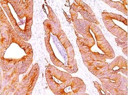
- Immunohistochemistry-Paraffin: Cytokeratin, pan Antibody (AE-1/AE-3) [NBP2-29429] - Analysis using Azide and BSA Free version of NBP2-29429. Human Colon Carcinoma stained with pan Cytokeratin Monoclonal Antibody cocktail (AE-1/AE3).

- Submitted by
- Novus Biologicals (provider)
- Main image

- Experimental details
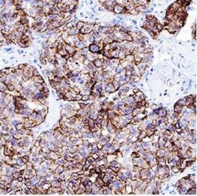
- Immunohistochemistry-Paraffin: Cytokeratin, pan Antibody (AE1 + AE3) [NBP2-29429] - Pan Cytokeratin was detected in immersion fixed paraffin-embedded sections of human liver cancer tissue using 1.7 ug/mL of mouse anti-pan Cytokeratin monoclonal (NBP2-29429, Novus Biologicals) for 1 hour at room temperature followed by anti-mouse IgG VisUCyte HRP polymer(VC001). Tissue was stained using DAB (brown) and counterstained with hematoxylin (blue).
Supportive validation
- Submitted by
- Novus Biologicals (provider)
- Main image
- Experimental details
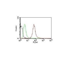
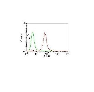
- Flow Cytometry: Cytokeratin, pan Antibody (AE1 + AE3) [NBP2-29429] - Human Pan-Cytokeratins on HeLa Cells. Black: Cells alone; Green: Isotype Control; Red: PE-labeled Pan-Cytokeratin Monoclonal Antibody (AE-1/AE-3)
- Submitted by
- Novus Biologicals (provider)
- Main image
- Experimental details
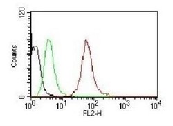
- Flow Cytometry: Cytokeratin, pan Antibody (AE1 + AE3) [NBP2-29429] - Analysis using Azide and BSA Free version of NBP2-29429. Black: Cells alone; Green: Isotype Control; Red: PE-labeled Pan-Cytokeratin Monoclonal Antibody (AE-1/AE-3).

- Submitted by
- Novus Biologicals (provider)
- Main image
- Experimental details
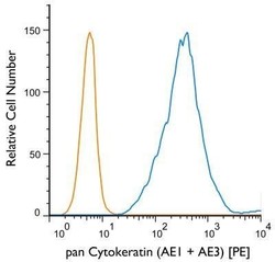
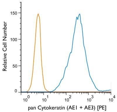
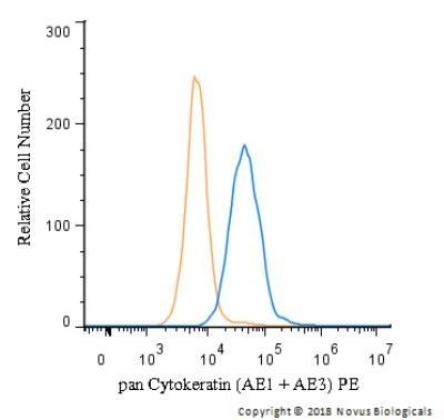
- Flow Cytometry: Cytokeratin, pan Antibody (AE1 + AE3) [NBP2-29429] - Using the PE direct conjugate An intracellular stain was performed on HT-29 cells with pan Cytokeratin (AE1 + AE3) antibody NBP2-33200PE (blue) and a matched isotype control NBP1-97005PE (orange). Cells were fixed with 4% PFA and then permeablized with 0.1% saponin. Cells were incubated in an antibody dilution of 2.5 ug/mL for 30 minutes at room temperature. Both antibodies were conjugated to Phycoerythrin.
- Submitted by
- Novus Biologicals (provider)
- Main image
- Experimental details
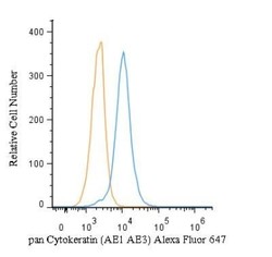
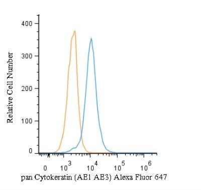
- Flow (Intracellular): Cytokeratin, pan Antibody (AE1 + AE3) [NBP2-29429] - An intracellular stain was performed on HeLa cells with pan Cytokeratin Antibody (AE1 + AE3) NBP2-33200AF647 (blue) and a matched isotype control (orange). Cells were fixed with 4% PFA and then permeabilized with 0.1% saponin. Cells were incubated in an antibody dilution of 5 ug/mL for 30 minutes at room temperature. Both antibodies were conjugated to Alexa Fluor 647. Image from the Alexa Fluor 647 version of this antibody.
- Submitted by
- Novus Biologicals (provider)
- Main image

- Experimental details
- Flow (Intracellular): Cytokeratin, pan Antibody (AE1 + AE3) [NBP2-29429] - An intracellular stain was performed on HeLa cells with pan Cytokeratin Antibody (AE1 + AE3) NBP2-33200PE (blue) and a matched isotype control (orange). Cells were fixed with 4% PFA and then permeabilized with 0.1% saponin. Cells were incubated in an antibody dilution of 2.5 ug/mL for 30 minutes at room temperature. Both antibodies were conjugated to phycoerythrin. Image from the PE version of this antibody.
- Submitted by
- Novus Biologicals (provider)
- Main image
- Experimental details
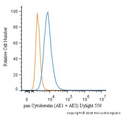
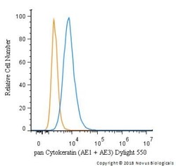
- Flow (Intracellular): Cytokeratin, pan Antibody (AE1 + AE3) [NBP2-29429] - An intracellular stain was performed on HeLa cells with pan Cytokeratin Antibody (AE1 + AE3) NBP2-33200R (blue) and a matched isotype control (orange). Cells were fixed with 4% PFA and then permeabilized with 0.1% saponin. Cells were incubated in an antibody dilution of 10 ug/mL for 30 minutes at room temperature. Both antibodies were conjugated to Dylight 550.