 Explore
 Validate
 Learn
Learn Western blot
 Immunohistochemistry
Antibody data
- Antibody Data
- Antigen structure
- References [3]
- Comments [0]
- Validations
- Immunohistochemistry [4]
- Other assay [1]
Submit
Validation data
Reference
Comment
Report error
- Product number
- MA1-06312 - Provider product page

- Provider
- Invitrogen Antibodies
- Product name
- Cytokeratin 1 Monoclonal Antibody (RCK103)
- Antibody type
- Monoclonal
- Antigen
- Other
- Description
- MA1-06312 detects basal cell cytokeratin in human, quail, chicken, rat, rabbit, hamster, canine, and guinea pig samples. MA1-06312 has sucessfully been used in Western blotting, flow cytometry, immunocytochemistry, and immunohistochemistry. By Western blotting, it detects a ~300 kDa protein representing basal cell cytokeratin. The MA1-06312 immunogen is a mix of cell preparations containing human cytokeratins.
- Reactivity
- Human, Rat, Canine, Chicken/Avian, Guinea Pig, Hamster, Porcine, Rabbit, Zebrafish
- Host
- Mouse
- Isotype
- IgG
- Antibody clone number
- RCK103
- Vial size
- 100 µg
- Concentration
- 1 mg/mL
- Storage
- Store at 4°C short term. For long term storage, store at -20°C, avoiding freeze/thaw cycles.
Submitted references Dynamic persistence of UPEC intracellular bacterial communities in a human bladder-chip model of urinary tract infection.
Loss of FOXC1 contributes to the corneal epithelial fate switch and pathogenesis.
Core transcription regulatory circuitry orchestrates corneal epithelial homeostasis.
Sharma K, Dhar N, Thacker VV, Simonet TM, Signorino-Gelo F, Knott GW, McKinney JD
eLife 2021 Jul 5;10
eLife 2021 Jul 5;10
Loss of FOXC1 contributes to the corneal epithelial fate switch and pathogenesis.
Li M, Zhu L, Liu J, Huang H, Guo H, Wang L, Li L, Gu S, Tan J, Zhong J, Wang B, Mao Z, Fan Y, Liu C, Yuan J, Ouyang H
Signal transduction and targeted therapy 2021 Jan 8;6(1):5
Signal transduction and targeted therapy 2021 Jan 8;6(1):5
Core transcription regulatory circuitry orchestrates corneal epithelial homeostasis.
Li M, Huang H, Li L, He C, Zhu L, Guo H, Wang L, Liu J, Wu S, Liu J, Xu T, Mao Z, Cao N, Zhang K, Lan F, Ding J, Yuan J, Liu Y, Ouyang H
Nature communications 2021 Jan 18;12(1):420
Nature communications 2021 Jan 18;12(1):420
No comments: Submit comment
Supportive validation
- Submitted by
- Invitrogen Antibodies (provider)
- Main image

- Experimental details
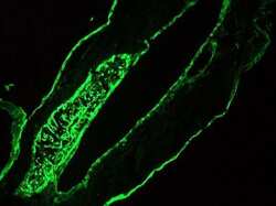
- Immunofluorescent analysis of 7 days old zebrafish embryo using Basal Cell Cytokeratin monoclonal antibody (Product # MA1-06312).

- Submitted by
- Invitrogen Antibodies (provider)
- Main image
- Experimental details
- Immunofluorescent analysis of 7 days old zebrafish embryo using Basal Cell Cytokeratin monoclonal antibody (Product # MA1-06312).

- Submitted by
- Invitrogen Antibodies (provider)
- Main image

- Experimental details
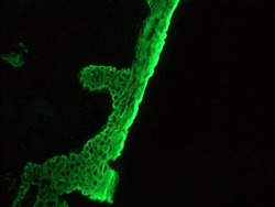
- Immunohistochemistry on frozen sections of human skin stained with Basal Cell Cytokeratin monoclonal antibody (Product # MA1-06312).

- Submitted by
- Invitrogen Antibodies (provider)
- Main image
- Experimental details
- Immunohistochemistry on frozen sections of human skin stained with Basal Cell Cytokeratin monoclonal antibody (Product # MA1-06312).

Supportive validation
- Submitted by
- Invitrogen Antibodies (provider)
- Main image
- Experimental details
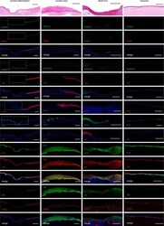
- Fig. 6 Pathological changes of corneal epithelium. H&E staining and immunofluorescence analysis of the indicated genes in corneal inflammatory (47 years old, female), ulcer (62 years old, female), alkali burn (33 years old, male), and leukoma (51 years old, female) tissues. Scale bars, 200 mum.
