Explore
 Validate
 Learn
Learn Western blot
 Flow cytometry
Antibody data
- Antibody Data
- Antigen structure
- References [1]
- Comments [0]
- Validations
- Flow cytometry [1]
Submit
Validation data
Reference
Comment
Report error
- Product number
- NBP2-33200AF647 - Provider product page

- Provider
- Novus Biologicals
- Product name
- Mouse Monoclonal Cytokeratin, pan Antibody
- Antibody type
- Monoclonal
- Description
- Protein A or G purified. Twenty human keratins are resolved with two-dimensional gel electrophoresis into acidic (pI 6.0) subfamilies. This antibody cocktail recognizes acidic (Type I or LMW) and basic (Type II or HMW) cytokeratins, which include CK1, CK3, CK4, CK5, CK6, CK8, CK10, CK14, CK15, CK16, and CK19. Many studies have shown the usefulness of keratins as markers in cancer research and tumor diagnosis. AE-1/AE-3 is a broad spectrum anti pan-cytokeratin antibody cocktail, which differentiates epithelial tumors from non-epithelial tumors e.g. squamous vs. adenocarcinoma of the lung, liver carcinoma, breast cancer, and esophageal cancer. It has been used to characterize the source of various neoplasms and to study the distribution of cytokeratin containing cells in epithelia during normal development and during the development of epithelial neoplasms. This antibody stains cytokeratins present in normal and abnormal human tissues and has shown high sensitivity in the recognition of epithelial cells and carcinomas.
- Reactivity
- Human, Mouse, Rat, Bovine, Canine, Chicken/Avian, Rabbit, Simian, Zebrafish
- Host
- Mouse
- Conjugate
- Red dye
- Isotype
- IgG
- Vial size
- 0.1 ml
- Storage
- Store at 4C in the dark.
Submitted references Cancer immunophenotyping by seven-colour multispectral imaging without tyramide signal amplification.
Ijsselsteijn ME, Brouwer TP, Abdulrahman Z, Reidy E, Ramalheiro A, Heeren AM, Vahrmeijer A, Jordanova ES, de Miranda NF
The journal of pathology. Clinical research 2019 Jan;5(1):3-11
The journal of pathology. Clinical research 2019 Jan;5(1):3-11
No comments: Submit comment
Supportive validation
- Submitted by
- Novus Biologicals (provider)
- Main image

- Experimental details
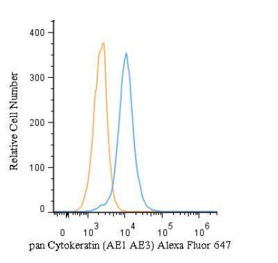
- Flow (Intracellular): pan Cytokeratin Antibody (AE1 + AE3) [Alexa Fluor 647] [NBP2-33200AF647] - An intracellular stain was performed on HeLa cells with pan Cytokeratin Antibody (AE1 + AE3) NBP2-33200AF647 (blue) and a matched isotype control (orange). Cells were fixed with 4% PFA and then permeabilized with 0.1% saponin. Cells were incubated in an antibody dilution of 5 ug/mL for 30 minutes at room temperature. Both antibodies were conjugated to Alexa Fluor 647.