Explore
 Validate
 Learn
Learn Western blot
 Immunohistochemistry
Antibody data
- Antibody Data
- Antigen structure
- References [4]
- Comments [0]
- Validations
- Immunohistochemistry [2]
- Flow cytometry [7]
Submit
Validation data
Reference
Comment
Report error
- Product number
- NBP2-33200 - Provider product page

- Provider
- Novus Biologicals
- Product name
- Mouse Monoclonal Cytokeratin, pan Antibody
- Antibody type
- Monoclonal
- Description
- Protein A or G purified. Twenty human keratins are resolved with two-dimensional gel electrophoresis into acidic (pI 6.0) subfamilies. This antibody cocktail recognizes acidic (Type I or LMW) and basic (Type II or HMW) cytokeratins, which include CK1, CK3, CK4, CK5, CK6, CK8, CK10, CK14, CK15, CK16, and CK19. Many studies have shown the usefulness of keratins as markers in cancer research and tumor diagnosis. AE-1/AE-3 is a broad spectrum anti pan-cytokeratin antibody cocktail, which differentiates epithelial tumors from non-epithelial tumors e.g. squamous vs. adenocarcinoma of the lung, liver carcinoma, breast cancer, and esophageal cancer. It has been used to characterize the source of various neoplasms and to study the distribution of cytokeratin containing cells in epithelia during normal development and during the development of epithelial neoplasms. This antibody stains cytokeratins present in normal and abnormal human tissues and has shown high sensitivity in the recognition of epithelial cells and carcinomas.
- Reactivity
- Human, Mouse, Rat, Bovine, Canine, Chicken/Avian, Rabbit, Simian, Zebrafish
- Host
- Mouse
- Isotype
- IgG
- Vial size
- 0.1 mg
- Concentration
- 1.0 mg/ml
- Storage
- Store at 4C short term. Aliquot and store at -20C long term. Avoid freeze-thaw cycles.
Submitted references Dysregulated Expression of the Nuclear Exosome Targeting Complex Component Rbm7 in Nonhematopoietic Cells Licenses the Development of Fibrosis.
Polyurethane scaffolds seeded with autologous cells can regenerate long esophageal gaps: An esophageal atresia treatment model.
Loss of atrx cooperates with p53-deficiency to promote the development of sarcomas and other malignancies.
Mad1 destabilizes p53 by preventing PML from sequestering MDM2.
Fukushima K, Satoh T, Sugihara F, Sato Y, Okamoto T, Mitsui Y, Yoshio S, Li S, Nojima S, Motooka D, Nakamura S, Kida H, Standley DM, Morii E, Kanto T, Yanagita M, Matsuura Y, Nagasawa T, Kumanogoh A, Akira S
Immunity 2020 Mar 17;52(3):542-556.e13
Immunity 2020 Mar 17;52(3):542-556.e13
Polyurethane scaffolds seeded with autologous cells can regenerate long esophageal gaps: An esophageal atresia treatment model.
Jensen T, Wanczyk H, Sharma I, Mitchell A, Sayej WN, Finck C
Journal of pediatric surgery 2019 Sep;54(9):1744-1754
Journal of pediatric surgery 2019 Sep;54(9):1744-1754
Loss of atrx cooperates with p53-deficiency to promote the development of sarcomas and other malignancies.
Oppel F, Tao T, Shi H, Ross KN, Zimmerman MW, He S, Tong G, Aster JC, Look AT
PLoS genetics 2019 Apr;15(4):e1008039
PLoS genetics 2019 Apr;15(4):e1008039
Mad1 destabilizes p53 by preventing PML from sequestering MDM2.
Wan J, Block S, Scribano CM, Thiry R, Esbona K, Audhya A, Weaver BA
Nature communications 2019 Apr 4;10(1):1540
Nature communications 2019 Apr 4;10(1):1540
No comments: Submit comment
Supportive validation
- Submitted by
- Novus Biologicals (provider)
- Main image
- Experimental details
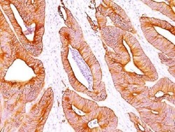
- Immunohistochemistry-Paraffin: Cytokeratin, pan Antibody (AE-1/AE-3) - Azide and BSA Free [NBP2-33200] - Analysis of Human Colon Carcinoma stained with pan Cytokeratin Monoclonal Antibody cocktail (AE-1/AE3).

- Submitted by
- Novus Biologicals (provider)
- Main image
- Experimental details
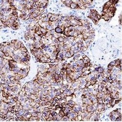
- Immunohistochemistry-Paraffin: Cytokeratin, pan Antibody (AE-1/AE-3) - Azide and BSA Free [NBP2-33200] - Pan Cytokeratin was detected in immersion fixed paraffin-embedded sections of human liver cancer tissue using 1.7 ug/mL of mouse anti-pan Cytokeratin monoclonal (NBP2-29429, Novus Biologicals) for 1 hour at room temperature followed by anti-mouse IgG VisU

Supportive validation
- Submitted by
- Novus Biologicals (provider)
- Main image

- Experimental details
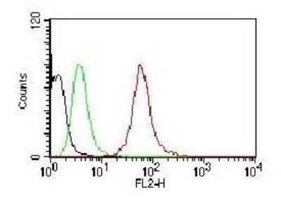
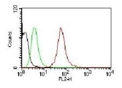
- Flow Cytometry: Cytokeratin, pan Antibody (AE-1/AE-3) - Azide and BSA Free [NBP2-33200] - Black: Cells alone; Green: Isotype Control; Red: PE-labeled Pan-Cytokeratin Monoclonal Antibody (AE-1/AE-3).
- Submitted by
- Novus Biologicals (provider)
- Main image
- Experimental details
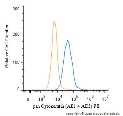
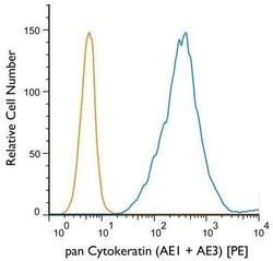
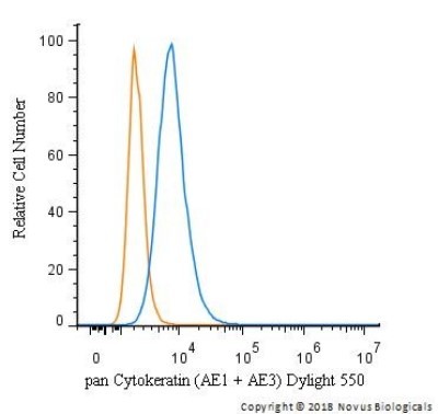
- Flow Cytometry: Cytokeratin, pan Antibody (AE-1/AE-3) - Azide and BSA Free [NBP2-33200] - Using the PE direct conjugate An intracellular stain was performed on HT-29 cells with pan Cytokeratin (AE1 + AE3) antibody NBP2-33200PE (blue) and a matched isotype control NBP1-97005PE (orange). Cells were fixed with 4% PFA and then permeablized with 0.1% saponin. Cells were incubated in an antibody dilution of 2.5 ug/mL for 30 minutes at room temperature. Both antibodies were conjugated to Phycoerythrin.

- Submitted by
- Novus Biologicals (provider)
- Main image

- Experimental details
- Flow Cytometry: Cytokeratin, pan Antibody (AE-1/AE-3) - Azide and BSA Free [NBP2-33200] - Analysis using Azide and BSA Free version of NBP2-29429. Black: Cells alone; Green: Isotype Control; Red: PE-labeled Pan-Cytokeratin Monoclonal Antibody (AE-1/AE-3).
- Submitted by
- Novus Biologicals (provider)
- Main image
- Experimental details
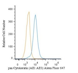
- Flow (Intracellular): Cytokeratin, pan Antibody (AE-1/AE-3) - Azide and BSA Free [NBP2-33200] - An intracellular stain was performed on HeLa cells with pan Cytokeratin Antibody (AE1 + AE3) NBP2-33200AF647 (blue) and a matched isotype control (orange). Cells were fixed with 4% PFA and then permeabilized with 0.1% saponin. Cells were incubated in an antibody dilution of 5 ug/mL for 30 minutes at room temperature. Both antibodies were conjugated to Alexa Fluor 647.
- Submitted by
- Novus Biologicals (provider)
- Main image
- Experimental details
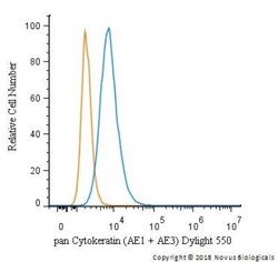
- Flow (Intracellular): Cytokeratin, pan Antibody (AE-1/AE-3) - Azide and BSA Free [NBP2-33200] - An intracellular stain was performed on HeLa cells with pan Cytokeratin Antibody (AE1 + AE3) NBP2-33200R (blue) and a matched isotype control (orange). Cells were fixed with 4% PFA and then permeabilized with 0.1% saponin. Cells were incubated in an antibody dilution of 10 ug/mL for 30 minutes at room temperature. Both antibodies were conjugated to Dylight 550.
- Submitted by
- Novus Biologicals (provider)
- Main image

- Experimental details
- Flow (Intracellular): Cytokeratin, pan Antibody (AE-1/AE-3) - Azide and BSA Free [NBP2-33200] - An intracellular stain was performed on HeLa cells with pan Cytokeratin [AE1 + AE3] Antibody NBP2-33200AF488 (blue) and a matched isotype control (orange). Cells were fixed with 4% PFA and then permeabilized with 0.1% saponin. Cells were incubated in an antibody dilution of 5 ug/mL for 30 minutes at room temperature. Both antibodies were conjugated to Alexa Fluor 488.

- Submitted by
- Novus Biologicals (provider)
- Main image

- Experimental details
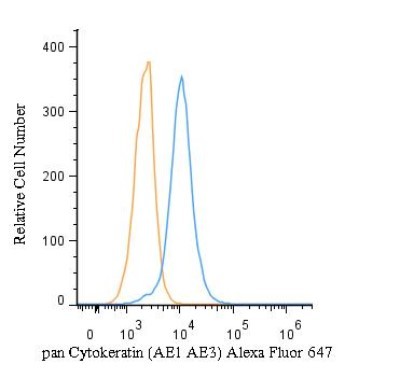
- Flow (Intracellular): Cytokeratin, pan Antibody (AE-1/AE-3) - Azide and BSA Free [NBP2-33200] - An intracellular stain was performed on HeLa cells with pan Cytokeratin Antibody (AE1 + AE3) NBP2-33200PE (blue) and a matched isotype control (orange). Cells were fixed with 4% PFA and then permeabilized with 0.1% saponin. Cells were incubated in an antibody dilution of 2.5 ug/mL for 30 minutes at room temperature. Both antibodies were conjugated to phycoerythrin. Image from the PE version of this antibody.