 Explore
 Validate
 Learn
Learn Western blot
 Immunohistochemistry
Antibody data
- Antibody Data
- Antigen structure
- References [0]
- Comments [0]
- Validations
- Immunohistochemistry [1]
- Flow cytometry [2]
Submit
Validation data
Reference
Comment
Report error
- Product number
- NBP2-34386-0.1mg - Provider product page

- Provider
- Novus Biologicals
- Product name
- Mouse Monoclonal Cytokeratin, pan Antibody
- Antibody type
- Monoclonal
- Description
- Protein G purified. This MAb recognizes cytokeratin 4, 5, 6, 8, 10, 13, and 18. This is a broad-spectrum antibody which has been reported to differentiate epithelial tumors from non-epithelial tumors. Many studies have shown the usefulness of keratins as markers in cancer research and tumor diagnosis.
- Reactivity
- Human, Mouse, Rat, Bovine, Goat, Guinea Pig, Porcine, Simian
- Host
- Mouse
- Isotype
- IgG
- Vial size
- 0.1 mg
- Concentration
- 0.2 mg/ml
- Storage
- Store at 4C.
No comments: Submit comment
Supportive validation
- Submitted by
- Novus Biologicals (provider)
- Main image

- Experimental details
- Immunohistochemistry-Paraffin: Cytokeratin, pan Antibody (SPM583) [NBP2-34386] - Formalin-fixed, paraffin-embedded colon (10X) stained with Cytokeratin, pan Antibody (SPM583).

Supportive validation
- Submitted by
- Novus Biologicals (provider)
- Main image

- Experimental details
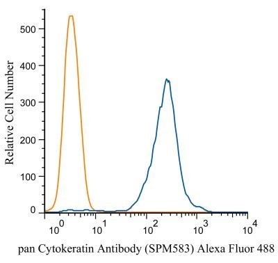
- Flow Cytometry: Cytokeratin, pan Antibody (SPM583) [NBP2-34386] - Analysis of Alexa Fluor (R) 488 conjugate of NBP2-34386. An intracellular stain was performed on HeLa cells with pan Cytokeratin (SMP583) antibody NBP2-34433AF488 (blue) and a matched isotype control NBP2-27287AF488 (orange). Cells were fixed with 4% PFA
- Submitted by
- Novus Biologicals (provider)
- Main image

- Experimental details
- Flow (Intracellular): Cytokeratin, pan Antibody (SPM583) [NBP2-34386] - Flow Cytometry: pan Cytokeratin Antibody (SPM583) - Azide and BSA Free [NBP2-34433] - An intracellular stain was performed on A549 cells with pan Cytokeratin Antibody (SPM583) NBP2-34433R (blue) and a matched isotype control (orange). Cells were fixed with 4% PFA and then permeabilized with 0.1% saponin. Cells were incubated in an antibody dilution of 5 ug/mL for 30 minutes at room temperature. Both antibodies were conjugated to Dylight 550. Image using the Azide and BSA Free form of this antibody.
