Explore
 Validate
 Learn
Learn Western blot
 Immunohistochemistry
Antibody data
- Antibody Data
- Antigen structure
- References [6]
- Comments [0]
- Validations
- Immunohistochemistry [1]
- Flow cytometry [8]
Submit
Validation data
Reference
Comment
Report error
- Product number
- NBP1-48348 - Provider product page

- Provider
- Novus Biologicals
- Proper citation
- Novus Cat#NBP1-48348, RRID:AB_10011106
- Product name
- Mouse Monoclonal Cytokeratin, pan Antibody
- Antibody type
- Monoclonal
- Description
- Protein A purified. This Mouse monoclonal Cytokeratin (Pan-reactive) antibody reacts with Cytokeratin peptides 4, 5, 6, 8, 10, 13, 18. Cytokeratins are a member of intermediate filaments subfamily represented in epithelial tissues.
- Reactivity
- Human, Mouse, Rat
- Host
- Mouse
- Isotype
- IgG
- Vial size
- 0.1 mg
- Concentration
- 1 mg/ml
- Storage
- Store at 4C. Do not freeze.
Submitted references Clinical Relevance of Immune Checkpoints on Circulating Tumor Cells in Breast Cancer.
Cancer immunophenotyping by seven-colour multispectral imaging without tyramide signal amplification.
In situ detection of CD73+ CD90+ CD105+ lineage: Mesenchymal stromal cells in human placenta and bone marrow specimens by chipcytometry.
Carotid artery stenosis with a high-intensity signal plaque on time-of-flight magnetic resonance angiography and association with evidence of intraplaque hypoxia.
Immune Defense Protein Expression in Highly Purified Mouse Lung Epithelial Cells.
Regulation of fibroblast lipid storage and myofibroblast phenotypes during alveolar septation in mice.
Papadaki MA, Koutsopoulos AV, Tsoulfas PG, Lagoudaki E, Aggouraki D, Monastirioti A, Koutoulaki C, Apostolopoulou CA, Merodoulaki AC, Papadaki C, Mavroudis D, Agelaki S
Cancers 2020 Feb 6;12(2)
Cancers 2020 Feb 6;12(2)
Cancer immunophenotyping by seven-colour multispectral imaging without tyramide signal amplification.
Ijsselsteijn ME, Brouwer TP, Abdulrahman Z, Reidy E, Ramalheiro A, Heeren AM, Vahrmeijer A, Jordanova ES, de Miranda NF
The journal of pathology. Clinical research 2019 Jan;5(1):3-11
The journal of pathology. Clinical research 2019 Jan;5(1):3-11
In situ detection of CD73+ CD90+ CD105+ lineage: Mesenchymal stromal cells in human placenta and bone marrow specimens by chipcytometry.
Consentius C, Mirenska A, Jurisch A, Reinke S, Scharm M, Zenclussen AC, Hennig C, Volk HD
Cytometry. Part A : the journal of the International Society for Analytical Cytology 2018 Jul;93(9):889-893
Cytometry. Part A : the journal of the International Society for Analytical Cytology 2018 Jul;93(9):889-893
Carotid artery stenosis with a high-intensity signal plaque on time-of-flight magnetic resonance angiography and association with evidence of intraplaque hypoxia.
Ogata A, Kawashima M, Wakamiya T, Nishihara M, Masuoka J, Nakahara Y, Ebashi R, Inoue K, Takase Y, Irie H, Abe T
Journal of neurosurgery 2017 Jun;126(6):1873-1878
Journal of neurosurgery 2017 Jun;126(6):1873-1878
Immune Defense Protein Expression in Highly Purified Mouse Lung Epithelial Cells.
Sinha M, Lowell CA
American journal of respiratory cell and molecular biology 2016 Jun;54(6):802-13
American journal of respiratory cell and molecular biology 2016 Jun;54(6):802-13
Regulation of fibroblast lipid storage and myofibroblast phenotypes during alveolar septation in mice.
McGowan SE, McCoy DM
American journal of physiology. Lung cellular and molecular physiology 2014 Oct 15;307(8):L618-31
American journal of physiology. Lung cellular and molecular physiology 2014 Oct 15;307(8):L618-31
No comments: Submit comment
Supportive validation
- Submitted by
- Novus Biologicals (provider)
- Main image

- Experimental details
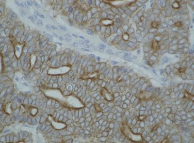
- Immunohistochemistry-Paraffin: Cytokeratin, pan Antibody (C-11) [NBP1-48348] - Analysis using the PE conjugate of NBP1-48348. Staining of cytokeratin on guinea pig breast carcinoma.
Supportive validation
- Submitted by
- Novus Biologicals (provider)
- Main image
- Experimental details
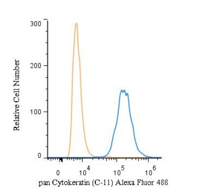
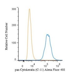
- Flow Cytometry: Cytokeratin, pan Antibody (C-11) [NBP1-48348] - An intracellular stain was performed on A549 cells with pan Cytokeratin Antibody (C-11) NBP1-48348F488 (blue) and a matched isotype control (orange). Cells were fixed with 4% PFA and then permeabilized with 0.1% saponin. Cells were incubated in an antibody dilution of 5 ug/mL for 30 minutes at room temperature. Both antibodies were conjugated to Alexa Fluor 488.
- Submitted by
- Novus Biologicals (provider)
- Main image
- Experimental details
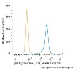
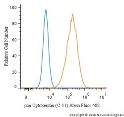
- Flow Cytometry: Cytokeratin, pan Antibody (C-11) [NBP1-48348] - An intracellular stain was performed on HeLa cells with pan Cytokeratin antibody (C-11) NBP1-48348AF488 (blue) and a matched isotype control (orange). Cells were fixed with 4% PFA and then permeablized with 0.1% saponin. Cells were incubated in an antibody dilution of 5 ug/mL for 30 minutes at room temperature. Both antibodies were conjugated to Alexa Fluor 488.

- Submitted by
- Novus Biologicals (provider)
- Main image
- Experimental details
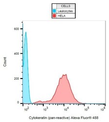
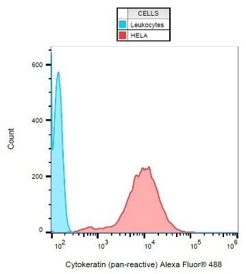
- Flow Cytometry: Cytokeratin, pan Antibody (C-11) [NBP1-48348] - Intracellular flow cytometry analysis of cytokeratin expression in HeLa cells.
- Submitted by
- Novus Biologicals (provider)
- Main image
- Experimental details
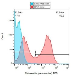
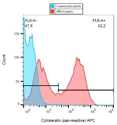
- Flow Cytometry: Cytokeratin, pan Antibody (C-11) [NBP1-48348] - Intracellular flow cytometry analysis of cytokeratin expression in HeLa cells using the APC conjugate.
- Submitted by
- Novus Biologicals (provider)
- Main image
- Experimental details
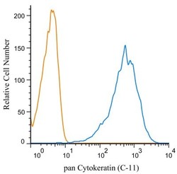
- Flow (Intracellular): Cytokeratin, pan Antibody (C-11) [NBP1-48348] - Intracellular flow cytometry analysis of cytokeratin expression in HT-29 human Caucasian colon adenocarcinoma cell line using anti-cytokeratin antibody (C-11) PE. Overlay with Isotype mouse IgG1 control. Image from the DyLight 594 version of this antibody.
- Submitted by
- Novus Biologicals (provider)
- Main image
- Experimental details
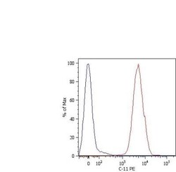
- Flow (Intracellular): Cytokeratin, pan Antibody (C-11) [NBP1-48348] - An intracellular stain was performed on HeLa cells with pan Cytokeratin (C-11) antibody NBP1-48348 (blue) and a matched isotype control NBP2-27287 (orange). Cells were fixed with 4% PFA and then permeablized with 0.1% saponin. Cells were incubated in an antibody dilution of 1 ug/mL for 30 minutes, followed by mouse F(ab)2 IgG (H+L) PE-conjugated secondary antibody (F0102B, R&D Systems).
- Submitted by
- Novus Biologicals (provider)
- Main image

- Experimental details
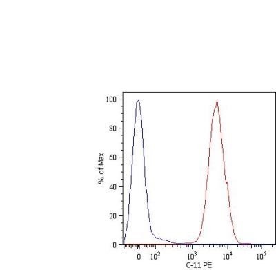
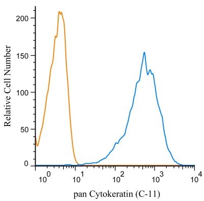
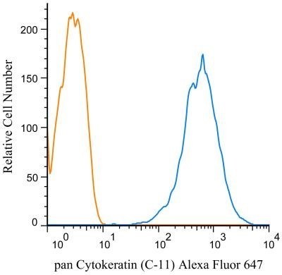
- Flow (Intracellular): Cytokeratin, pan Antibody (C-11) [NBP1-48348] - An intracellular stain was performed on HeLa cells with pan Cytokeratin antibody (C-11) NBP1-48348AF647 (blue) and a matched isotype control NBP2-27287AF647 (orange). Cells were fixed with 4% PFA and then permeablized with 0.1% saponin. Cells were incubated in an antibody dilution of 2 ug/mL for 30 minutes at room temperature. Both antibodies were conjugated to Alexa Fluor 647.
- Submitted by
- Novus Biologicals (provider)
- Main image
- Experimental details
- Flow (Intracellular): Cytokeratin, pan Antibody (C-11) [NBP1-48348] - An intracellular stain was performed on HepG2 cells with pan Cytokeratin Antibody (C-11) NBP1-48348F488 (blue) and a matched isotype control (orange). Cells were fixed with 4% PFA and then permeabilized with 0.1% saponin. Cells were incubated in an antibody dilution of 10 ug/mL for 30 minutes at room temperature. Both antibodies were conjugated to Alexa Fluor 488.