 Explore
 Validate
 Learn
Learn Western blot
 Immunocytochemistry
Antibody data
- Antibody Data
- Antigen structure
- References [12]
- Comments [0]
- Validations
- Western blot [1]
- Immunohistochemistry [2]
Submit
Validation data
Reference
Comment
Report error
- Product number
- NB100-2756 - Provider product page

- Provider
- Novus Biologicals
- Proper citation
- Novus Cat#NB100-2756, RRID:AB_10001893
- Product name
- Mouse Monoclonal Cytokeratin 1 Antibody
- Antibody type
- Monoclonal
- Description
- Protein G purified.
- Reactivity
- Human, Rat
- Host
- Mouse
- Isotype
- IgG
- Vial size
- 0.1 ml
- Concentration
- 1.0 mg/ml
- Storage
- Store at 4C short term. Aliquot and store at -20C long term. Avoid freeze-thaw cycles.
Submitted references The exosomal integrin α5β1/AEP complex derived from epithelial ovarian cancer cells promotes peritoneal metastasis through regulating mesothelial cell proliferation and migration.
Peptidylarginine deiminase is involved in maintaining the cornified oral mucosa of rats.
Phosphodiesterase 4B plays a role in benzophenone-3-induced phototoxicity in normal human keratinocytes.
Attenuated kallikrein-related peptidase activity disrupts desquamation and leads to stratum corneum thickening in human skin equivalent models.
Blocking glutamate carboxypeptidase II inhibits glutamate excitotoxicity and regulates immune responses in experimental autoimmune encephalomyelitis.
A new model for preclinical testing of dermal substitutes for human skin reconstruction.
Human amniotic fluid derived cells can competently substitute dermal fibroblasts in a tissue-engineered dermo-epidermal skin analog.
Dysregulation of the repressive H3K27 trimethylation mark in head and neck squamous cell carcinoma contributes to dysregulated squamous differentiation.
Modified plastic compression of collagen hydrogels provides an ideal matrix for clinically applicable skin substitutes.
A human keratin 10 knockout causes recessive epidermolytic hyperkeratosis.
Characterization and comparison of reconstructed skin models: morphological and immunohistochemical evaluation.
Activated keratinocytes in the epidermis of hypertrophic scars.
Li X, Tang M, Zhu Q, Wang X, Lin Y, Wang X
Cellular oncology (Dordrecht) 2020 Apr;43(2):263-277
Cellular oncology (Dordrecht) 2020 Apr;43(2):263-277
Peptidylarginine deiminase is involved in maintaining the cornified oral mucosa of rats.
Arita S, Hatta M, Uchida K, Kita T, Okamura K, Ryu T, Murakami H, Sakagami R, Yamazaki J
Journal of periodontal research 2018 Oct;53(5):750-761
Journal of periodontal research 2018 Oct;53(5):750-761
Phosphodiesterase 4B plays a role in benzophenone-3-induced phototoxicity in normal human keratinocytes.
Kim HJ, Lee E, Lee M, Ahn S, Kim J, Liu J, Jin SH, Ha J, Bae IH, Lee TR, Noh M
Toxicology and applied pharmacology 2018 Jan 1;338:174-181
Toxicology and applied pharmacology 2018 Jan 1;338:174-181
Attenuated kallikrein-related peptidase activity disrupts desquamation and leads to stratum corneum thickening in human skin equivalent models.
McGovern JA, Meinert C, de Veer SJ, Hollier BG, Parker TJ, Upton Z
The British journal of dermatology 2017 Jan;176(1):145-158
The British journal of dermatology 2017 Jan;176(1):145-158
Blocking glutamate carboxypeptidase II inhibits glutamate excitotoxicity and regulates immune responses in experimental autoimmune encephalomyelitis.
Ha D, Bing SJ, Ahn G, Kim J, Cho J, Kim A, Herath KH, Yu HS, Jo SA, Cho IH, Jee Y
The FEBS journal 2016 Sep;283(18):3438-56
The FEBS journal 2016 Sep;283(18):3438-56
A new model for preclinical testing of dermal substitutes for human skin reconstruction.
Hartmann-Fritsch F, Biedermann T, Braziulis E, Meuli M, Reichmann E
Pediatric surgery international 2013 May;29(5):479-88
Pediatric surgery international 2013 May;29(5):479-88
Human amniotic fluid derived cells can competently substitute dermal fibroblasts in a tissue-engineered dermo-epidermal skin analog.
Hartmann-Fritsch F, Hosper N, Luginbühl J, Biedermann T, Reichmann E, Meuli M
Pediatric surgery international 2013 Jan;29(1):61-9
Pediatric surgery international 2013 Jan;29(1):61-9
Dysregulation of the repressive H3K27 trimethylation mark in head and neck squamous cell carcinoma contributes to dysregulated squamous differentiation.
Gannon OM, Merida de Long L, Endo-Munoz L, Hazar-Rethinam M, Saunders NA
Clinical cancer research : an official journal of the American Association for Cancer Research 2013 Jan 15;19(2):428-41
Clinical cancer research : an official journal of the American Association for Cancer Research 2013 Jan 15;19(2):428-41
Modified plastic compression of collagen hydrogels provides an ideal matrix for clinically applicable skin substitutes.
Braziulis E, Diezi M, Biedermann T, Pontiggia L, Schmucki M, Hartmann-Fritsch F, Luginbühl J, Schiestl C, Meuli M, Reichmann E
Tissue engineering. Part C, Methods 2012 Jun;18(6):464-74
Tissue engineering. Part C, Methods 2012 Jun;18(6):464-74
A human keratin 10 knockout causes recessive epidermolytic hyperkeratosis.
Müller FB, Huber M, Kinaciyan T, Hausser I, Schaffrath C, Krieg T, Hohl D, Korge BP, Arin MJ
Human molecular genetics 2006 Apr 1;15(7):1133-41
Human molecular genetics 2006 Apr 1;15(7):1133-41
Characterization and comparison of reconstructed skin models: morphological and immunohistochemical evaluation.
Boelsma E, Gibbs S, Faller C, Ponec M
Acta dermato-venereologica 2000 Mar-Apr;80(2):82-8
Acta dermato-venereologica 2000 Mar-Apr;80(2):82-8
Activated keratinocytes in the epidermis of hypertrophic scars.
Machesney M, Tidman N, Waseem A, Kirby L, Leigh I
The American journal of pathology 1998 May;152(5):1133-41
The American journal of pathology 1998 May;152(5):1133-41
No comments: Submit comment
Supportive validation
- Submitted by
- Novus Biologicals (provider)
- Main image
- Experimental details
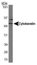
- Western Blot: Cytokeratin 1 Antibody (LHK1) [NB100-2756] - Analysis of Cytokeratin 1 in Caco-2.

Supportive validation
- Submitted by
- Novus Biologicals (provider)
- Main image
- Experimental details
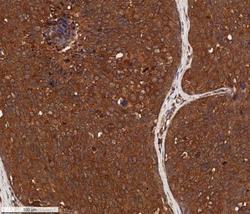
- Immunohistochemistry-Paraffin: Cytokeratin 1 Antibody (LHK1) [NB100-2756] - IHC analysis of a formalin fixed paraffin-embedded (FFPE) human breast cancer using 5ug/ml conc. of Cytokeratin 1 antibody (clone LHK1) on a Bond Rx autostainer (Leica Biosystems). The assay involved 20 minutes of heat induced antigen retrieval (HIER) using 10mM sodium citrate buffer (pH 6.0) and endogenous peroxidase quenching with peroxide block. The sections were incubated with primary antibody for 30 minutes and Bond Polymer Refine Detection (Leica Biosystems) with DAB was used for signal development followed by counterstaining with hematoxylin. Whole slide scanning and capturing of representative images (20X) was performed using Aperio AT2 (Leica Biosystems). A membrane-cytoplasmic staining of Cytokeratin 1 was observed in the cancer cells with signal localized more to the cell membranes. Tumor cores showed a decreased staining and tumor stroma was mostly negative for Cytokeratin 1.

- Submitted by
- Novus Biologicals (provider)
- Main image

- Experimental details
- Immunohistochemistry-Paraffin: Cytokeratin 1 Antibody (LHK1) [NB100-2756] - IHC analysis of a formalin fixed paraffin-embedded (FFPE) human breast cancer using 5ug/ml conc. of Cytokeratin 1 antibody (clone LHK1) on a Bond Rx autostainer (Leica Biosystems). The assay involved 20 minutes of heat induced antigen retrieval (HIER) using 10mM sodium citrate buffer (pH 6.0) and endogenous peroxidase quenching with peroxide block. The sections were incubated with primary antibody for 30 minutes and Bond Polymer Refine Detection (Leica Biosystems) with DAB was used for signal development followed by counterstaining with hematoxylin. Whole slide scanning and capturing of representative images (20X) was performed using Aperio AT2 (Leica Biosystems). A membrane-cytoplasmic staining of Cytokeratin 1 was observed in the cancer cells with signal localized more to the cell membranes. Tumor stroma was mostly negative for Cytokeratin 1. Staining was performed by Histowiz.
