 Explore
 Validate
 Learn
Learn Western blot
Antibody data
- Antibody Data
- Antigen structure
- References [1]
- Comments [0]
- Validations
- Western blot [1]
- Immunocytochemistry [1]
- Immunohistochemistry [2]
Submit
Validation data
Reference
Comment
Report error
- Product number
- MA5-16032 - Provider product page

- Provider
- Invitrogen Antibodies
- Product name
- Cytokeratin 1 Monoclonal Antibody (LHK1)
- Antibody type
- Monoclonal
- Antigen
- Synthetic peptide
- Reactivity
- Human, Rat
- Host
- Mouse
- Isotype
- IgG
- Antibody clone number
- LHK1
- Vial size
- 100 μL
- Concentration
- 1.0 mg/mL
- Storage
- Store at 4°C short term. For long term storage, store at -20°C, avoiding freeze/thaw cycles.
Submitted references Transcriptional Mechanisms of Resistance to Anti-PD-1 Therapy.
Ascierto ML, Makohon-Moore A, Lipson EJ, Taube JM, McMiller TL, Berger AE, Fan J, Kaunitz GJ, Cottrell TR, Kohutek ZA, Favorov A, Makarov V, Riaz N, Chan TA, Cope L, Hruban RH, Pardoll DM, Taylor BS, Solit DB, Iacobuzio-Donahue CA, Topalian SL
Clinical cancer research : an official journal of the American Association for Cancer Research 2017 Jun 15;23(12):3168-3180
Clinical cancer research : an official journal of the American Association for Cancer Research 2017 Jun 15;23(12):3168-3180
No comments: Submit comment
Supportive validation
- Submitted by
- Invitrogen Antibodies (provider)
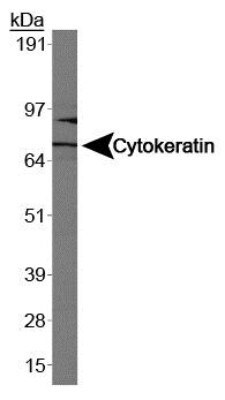
- Main image

- Experimental details
- Western blot analysis of Cytokeratin 1 in Caco-2. Sample was incubated in Cytokeratin 1 monoclonal antibody (Product # MA5-16032).
Supportive validation
- Submitted by
- Invitrogen Antibodies (provider)
- Main image
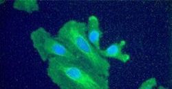
- Experimental details
- Immunofluorescent analysis of Cytokeratin 1 using a monoclonal antibody (Product # MA5-16032).

Supportive validation
- Submitted by
- Invitrogen Antibodies (provider)
- Main image
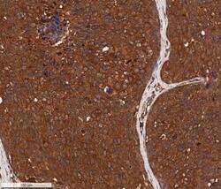
- Experimental details
- Immunohistochemical analysis of Cytokeratin 1 in formalin fixed paraffin-embedded (FFPE) human breast cancer. Samples were incubated in Cytokeratin 1 monoclonal antibody (Product # MA5-16032) using a dilution of 5 µg/mL. Bond Rx autostainer (Leica Biosystems). The assay involved 20 minutes of heat induced antigen retrieval (HIER) using 10mM sodium citrate buffer (pH 6.0) and endogenous peroxidase quenching with peroxide block. The sections were incubated with primary antibody for 30 minutes and Bond Polymer Refine Detection (Leica Biosystems) with DAB was used for signal development followed by counterstaining with hematoxylin. Whole slide scanning and capturing of representative images (20X) was performed using Aperio AT2 (Leica Biosystems). A membrane-cytoplasmic staining of Cytokeratin 1 was observed in the cancer cells with signal localized more to the cell membranes. Tumor cores showed a decreased staining and tumor stroma was mostly negative for Cytokeratin 1.

- Submitted by
- Invitrogen Antibodies (provider)
- Main image
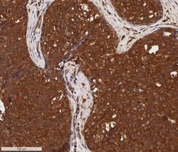
- Experimental details
- Immunohistochemical analysis of Cytokeratin 1 in formalin fixed paraffin-embedded (FFPE) human breast cancer. Samples were incubated in Cytokeratin 1 monoclonal antibody (Product # MA5-16032) using a dilution of 5 µg/mL. Bond Rx autostainer (Leica Biosystems). The assay involved 20 minutes of heat induced antigen retrieval (HIER) using 10mM sodium citrate buffer (pH 6.0) and endogenous peroxidase quenching with peroxide block. The sections were incubated with primary antibody for 30 minutes and Bond Polymer Refine Detection (Leica Biosystems) with DAB was used for signal development followed by counterstaining with hematoxylin. Whole slide scanning and capturing of representative images (20X) was performed using Aperio AT2 (Leica Biosystems). A membrane-cytoplasmic staining of Cytokeratin 1 was observed in the cancer cells with signal localized more to the cell membranes. Tumor stroma was mostly negative for Cytokeratin 1. Staining was performed by Histowiz.
