 Explore
 Validate
 Learn
Learn Western blot
 Immunocytochemistry
Antibody data
- Antibody Data
- Antigen structure
- References [1]
- Comments [0]
- Validations
- Western blot [1]
- Immunohistochemistry [3]
Submit
Validation data
Reference
Comment
Report error
- Product number
- GTX82905 - Provider product page

- Provider
- GeneTex
- Proper citation
- GeneTex Cat#GTX82905, RRID:AB_625814
- Product name
- nNOS antibody
- Antibody type
- Polyclonal
- Reactivity
- Human, Mouse, Rat, Bovine, Guinea Pig, Rabbit, Zebrafish
- Host
- Rabbit
Submitted references The effect of sumatriptan on nitric oxide synthase enzyme production after iatrogenic inflammation in the brain stem of adolescent rats: A randomized, controlled, experimental study.
Demirpence S, Kurul SH, Kiray M, Tugyan K, Yilmaz O, Köse G
Current therapeutic research, clinical and experimental 2009 Apr;70(2):129-35
Current therapeutic research, clinical and experimental 2009 Apr;70(2):129-35
No comments: Submit comment
Supportive validation
- Submitted by
- GeneTex (provider)
- Main image
- Experimental details
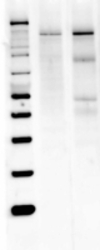
- Western blot analysis of nNOS was performed by loading 40 ?g of Mouse (Lane 1) and Rat Brain (Lane 2) tissue lysate onto a 4-12% Bis-Tris polyacrylamide gel. Proteins were transferred to a Nitrocellulose membrane. Membranes were probed with a rabbit polyclonal antibody (GTX82905) recognizing nNOS at a dilution of 1:1000.

Supportive validation
- Submitted by
- GeneTex (provider)
- Main image
- Experimental details
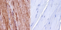
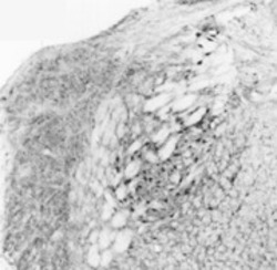
- Immunohistochemistry was performed on normal biopsies of deparaffinized mouse skeletal muscle tissue. To expose target proteins, heat induced antigen retrieval was performed using 10mM sodium citrate (pH 6.0) buffer, microwaved for 8-15 minutes. Following antigen retrieval tissues were blocked in 3% BSA-PBS for 30 minutes at room temperature. Tissues were then probed at a dilution of 1:400 with a Rabbit Polyclonal Antibody recognizing nNOS (GTX82905) or without primary antibody (negative control) overnight at 4¢XC in a humidified chamber. Tissues were washed extensively with PBST and endogenous peroxidase activity was quenched with a peroxidase suppressor. Detection was performed using a biotin-conjugated secondary antibody and SA-HRP, followed by colorimetric detection using DAB. Tissues were counterstained with hematoxylin and prepped for mounting.
- Submitted by
- GeneTex (provider)
- Main image
- Experimental details
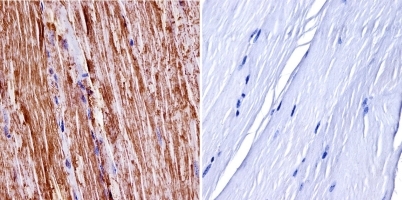
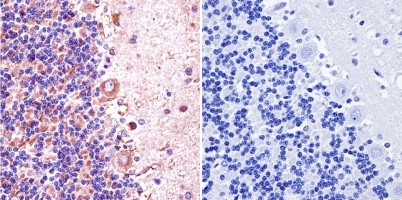
- Immunohistochemistry was performed on normal biopsies of deparaffinized Rat cerebellum tissue. To expose target proteins, heat induced antigen retrieval was performed using 10mM sodium citrate (pH 6.0) buffer, microwaved for 8-15 minutes. Following antigen retrieval tissues were blocked in 3% BSA-PBS for 30 minutes at room temperature. Tissues were then probed at a dilution of 1:200 with a Rabbit Polyclonal Antibody recognizing nNOS or without primary antibody (negative control) overnight at 4¢XC in a humidified chamber. Tissues were washed extensively with PBST and endogenous peroxidase activity was quenched with a peroxidase suppressor. Detection was performed using a biotin-conjugated secondary antibody and SA-HRP, followed by colorimetric detection using DAB. Tissues were counterstained with hematoxylin and prepped for mounting.
- Submitted by
- GeneTex (provider)
- Main image
- Experimental details
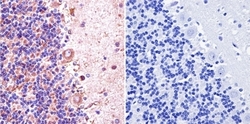
- Immunohistochemical staining of bNOS in rat brain using GTX82905.
