Explore
 Validate
 Learn
Learn Western blot
Antibody data
- Antibody Data
- Antigen structure
- References [0]
- Comments [0]
- Validations
- Western blot [2]
- Immunocytochemistry [1]
- Immunohistochemistry [1]
Submit
Validation data
Reference
Comment
Report error
- Product number
- SP5116P - Provider product page

- Provider
- Acris Antibodies GmbH
- Proper citation
- Acris Antibodies GmbH Cat#SP5116P, RRID:AB_1001073
- Product name
- anti NOS1
- Antibody type
- Polyclonal
- Antigen
- Synthetic peptide corresponding to residues C N(1411) R L R S E S I A F I E E S K(1425) of human nNOS.
- Reactivity
- Human, Mouse, Rat
- Host
- Rabbit
- Vial size
- 0.1 mg
- Concentration
- 1.0 mg/ml
No comments: Submit comment
Supportive validation
- Submitted by
- Acris Antibodies GmbH (provider)
- Main image
- Experimental details
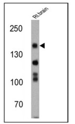
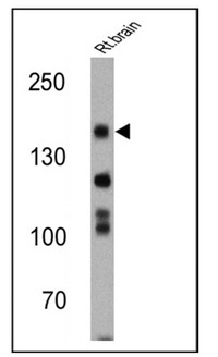
- Western blot analysis of nNOS was performed by loading 25 ug of rat brain cell lysates onto an SDS polyacrylamide gel. Proteins were transferred to a PVDF membrane and blocked at 4ºC overnight. The membrane was probed with a nNOS polyclonal antibody (Cat.-No SP5116P) at a dilution of 1:200 overnight at 4°C, washed in TBST, and probed with an HRP-conjugated secondary antibody for 1 hr at room temperature in the dark. Chemiluminescent detection was performed using Pierce ECL Plus Western Blotting Substrate. Results show a band at approx. 160 kDa.
- Submitted by
- Acris Antibodies GmbH (provider)
- Main image
- Experimental details
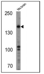
- Western blot analysis of nNOS was performed by loading 25 ug of mouse brain cell lysates onto an SDS polyacrylamide gel. Proteins were transferred to a PVDF membrane and blocked at 4ºC overnight. The membrane was probed with a nNOS polyclonal antibody (Cat.-No SP5116P) at a dilution of 1:500 overnight at 4°C, washed in TBST, and probed with an HRP-conjugated secondary antibody for 1 hr at room temperature in the dark. Chemiluminescent detection was performed using Pierce ECL Plus Western Blotting Substrate. Results show a band at approx. 160 kDa.

Supportive validation
- Submitted by
- Acris Antibodies GmbH (provider)
- Main image
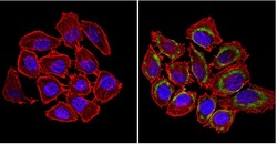
- Experimental details
- Immunofluorescent analysis of nNOS (green) showing staining in the cytoplasm and membrane of U251 cells (right) compared to a negative control without primary antibody (left). Formalin-fixed cells were permeabilized with 0.1% Triton X-100 in TBS for 5-10 minutes and blocked with 3% BSA-PBS for 30 minutes at room temperature. Cells were probed with an nNOS polyclonal antibody (Cat.-No SP5116P) in 3% BSA-PBS at a dilution of 1:100 and incubated overnight at 4ºC in a humidified chamber. Cells were washed with PBST and incubated with a DyLight-conjugated secondary antibody in PBS at room temperature in the dark. F-Actin (red) was stained with a fluorescent red phalloidin and nuclei (blue) were stained with Hoechst or DAPI. Images were taken at a magnification of 60x.

Supportive validation
- Submitted by
- Acris Antibodies GmbH (provider)
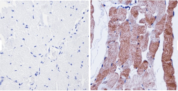
- Main image
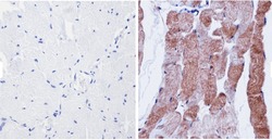
- Experimental details
- Immunohistochemistry analysis of nNOS showing positive staining in the cytoplasm of paraffin-treated Human skeletal muscle (right) compared with a negative control in the absence of primary antibody (left). To expose target proteins, antigen retrieval method was performed using 10mM sodium citrate (pH 6.0) microwaved for 8-15 min. Following antigen retrieval, tissues were blocked in 3% H2O2-methanol for 15 min at room temperature, washed with ddH2O and PBS, and then probed with a nNOS polyclonal antibody (Cat.-No SP5116P) diluted by 3% BSA-PBS at a dilution of 1:200 overnight at 4°C in a humidified chamber. Tissues were washed extensively PBST and detection was performed using an HRP-conjugated secondary antibody followed by colorimetric detection using a DAB kit. Tissues were counterstained with hematoxylin and dehydrated with ethanol and xylene to prep for mounting.