Explore
 Validate
 Learn
Learn Western blot
Antibody data
- Antibody Data
- Antigen structure
- References [4]
- Comments [0]
- Validations
- Western blot [7]
- Immunohistochemistry [3]
- Other assay [2]
Submit
Validation data
Reference
Comment
Report error
- Product number
- PA5-35879 - Provider product page

- Provider
- Invitrogen Antibodies
- Product name
- Phospho-eNOS (Ser1177) Polyclonal Antibody
- Antibody type
- Polyclonal
- Antigen
- Synthetic peptide
- Description
- Recommended positive controls: Raji, NCI-H929, THP-1, THP-1 (100 nM PMA treatment for 6, 24, 48 hr).
- Concentration
- 0.94 mg/mL
Submitted references Global REACH 2018: dysfunctional extracellular microvesicles in Andean highlander males with excessive erythrocytosis.
Impaired TRPV4-eNOS signaling in trabecular meshwork elevates intraocular pressure in glaucoma.
Endothelin-1-induced endothelial microvesicles impair endothelial cell function.
Effects of circulating extracellular microvesicles from spinal cord-injured adults on endothelial cell function.
Brewster LM, Bain AR, Garcia VP, Fandl HK, Stone R, DeSouza NM, Greiner JJ, Tymko MM, Vizcardo-Galindo GA, Figueroa-Mujica RJ, Villafuerte FC, Ainslie PN, DeSouza CA
American journal of physiology. Heart and circulatory physiology 2021 May 1;320(5):H1851-H1861
American journal of physiology. Heart and circulatory physiology 2021 May 1;320(5):H1851-H1861
Impaired TRPV4-eNOS signaling in trabecular meshwork elevates intraocular pressure in glaucoma.
Patel PD, Chen YL, Kasetti RB, Maddineni P, Mayhew W, Millar JC, Ellis DZ, Sonkusare SK, Zode GS
Proceedings of the National Academy of Sciences of the United States of America 2021 Apr 20;118(16)
Proceedings of the National Academy of Sciences of the United States of America 2021 Apr 20;118(16)
Endothelin-1-induced endothelial microvesicles impair endothelial cell function.
Brewster LM, Garcia VP, Levy MV, Stockelman KA, Goulding A, DeSouza NM, Greiner JJ, Hijmans JG, DeSouza CA
Journal of applied physiology (Bethesda, Md. : 1985) 2020 Jun 1;128(6):1497-1505
Journal of applied physiology (Bethesda, Md. : 1985) 2020 Jun 1;128(6):1497-1505
Effects of circulating extracellular microvesicles from spinal cord-injured adults on endothelial cell function.
Brewster LM, Coombs GB, Garcia VP, Hijmans JG, DeSouza NM, Stockelman KA, Barak OF, Mijacika T, Dujic Z, Greiner JJ, Phillips AA, Ainslie PN, DeSouza CA
Clinical science (London, England : 1979) 2020 Apr 17;134(7):777-789
Clinical science (London, England : 1979) 2020 Apr 17;134(7):777-789
No comments: Submit comment
Supportive validation
- Submitted by
- Invitrogen Antibodies (provider)
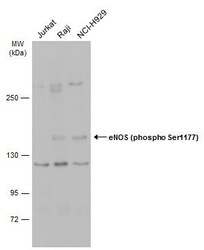
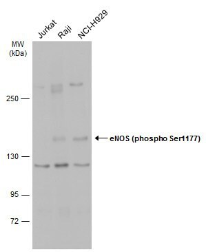
- Main image

- Experimental details
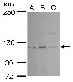
- Western blot analysis of Phospho-eNOS pSer1177 using A) 30 µg Jurkat whole cell lysate (B) 30 µg Raji whole cell lysate and C) 30 µg NCI-H929 whole cell lysate. Samples were loaded onto a 5% SDS-PAGE gel and probed with a Phospho-eNOS pSer1177 polyclonal antibody (Product # PA5-35879) at a dilution of 1:500.
- Submitted by
- Invitrogen Antibodies (provider)
- Main image

- Experimental details
- Western blot analysis of Phospho-eNOS pSer1177 using A) 30 µg THP-1 whole cell lysate (untreated) (B) 30 µg THP-1 whole cell lysate (100 nM PMA treatment for 6 hr) (C) 30 µg THP-1 whole cell lysate (untreated) (D) 30 µg THP-1 whole cell lysate (100 nM PMA treatment for 24 hr) (E) 30 µg THP-1 whole cell lysate (untreated) and F) 30 µg THP-1 whole cell lysate (100 nM PMA treatment for 48 hr). Samples were loaded onto a 5% SDS-PAGE gel and probed with a Phospho-eNOS pSer1177 polyclonal antibody (Product # PA5-35879) at a dilution of 1:500.

- Submitted by
- Invitrogen Antibodies (provider)
- Main image
- Experimental details
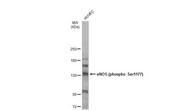
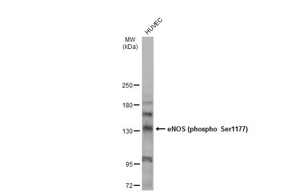
- Western Blot using Phospho-eNOS (Ser1177) Polyclonal Antibody (Product # PA5-35879). Whole cell extract (50 µg) was separated by 5% SDS-PAGE, and the membrane was blotted with Phospho-eNOS (Ser1177) Polyclonal Antibody (Product # PA5-35879) diluted at 1:1,000. The HRP-conjugated anti-rabbit IgG antibody was used to detect the primary antibody.
- Submitted by
- Invitrogen Antibodies (provider)
- Main image
- Experimental details
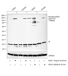
- Western blot was performed using Anti-Phospho-eNOS (Ser1177) Polyclonal Antibody (Product # PA5-35879) and a 160 kDa band corresponding to Phospho-eNOS (Ser1177) was observed to be upregulated upon treatment of HAEC and HUVEC with VEGF (10 ng/mL for 60 min) and H2O2 (300 µM for 30 min). Whole cell extracts (30 µg lysate) of HAEC (Lane 1), HAEC treated with VEGF (10 ng/mL for 60 min) (Lane 2), HUVEC (Lane 3), HUVEC treated with VEGF (10 ng/mL for 60 min) (Lane 4), HAEC (Lane 5), HAEC treated with H2O2 (300 µM for 30 min) (Lane 6), HUVEC (Lane 7), HUVEC treated with H2O2 (300 µM for 30 min) (Lane 8) were electrophoresed using NuPAGE™ 4-12% Bis-Tris Protein Gel (Product # NP0322BOX). Resolved proteins were then transferred onto a nitrocellulose membrane (Product # IB23001) by iBlot® 2 Dry Blotting System (Product # IB21001). The blot was probed with the primary antibody (1:1000 dilution) and detected by chemiluminescence with Goat anti-Rabbit IgG (H+L) Superclonal™ Recombinant Secondary Antibody, HRP (Product # A27036,1:20000 dilution) using the iBright™ FL1500 Imaging System (Product # A44115). Chemiluminescent detection was performed using SuperSignal™ West Pico PLUS Chemiluminescent Substrate (Product # 34580). An uncharacterized band (*) was observed at ~20 kDa.

- Submitted by
- Invitrogen Antibodies (provider)
- Main image

- Experimental details
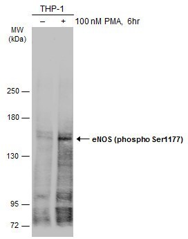
- Western Blot using Phospho-eNOS (Ser1177) Polyclonal Antibody (Product # PA5-35879). Untreated (–) and treated (+) THP-1 whole cell extracts (30 µg) were separated by 5% SDS-PAGE, and the membrane was blotted with Phospho-eNOS (Ser1177) Polyclonal Antibody (Product # PA5-35879) diluted at 1:500. The HRP-conjugated anti-rabbit IgG antibody was used to detect the primary antibody.
- Submitted by
- Invitrogen Antibodies (provider)
- Main image
- Experimental details
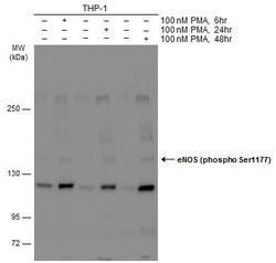
- Western Blot analysis of Phospho-eNOS (Ser1177) was performed by separating 30 µg of untreated (–) and treated (+) THP-1 whole cell extracts by 5% SDS-PAGE. Proteins were transferred to a membrane and probed with a Phospho-eNOS (Ser1177) Polyclonal Antibody (Product # PA5-35879) at a dilution of 1:500.
- Submitted by
- Invitrogen Antibodies (provider)
- Main image
- Experimental details
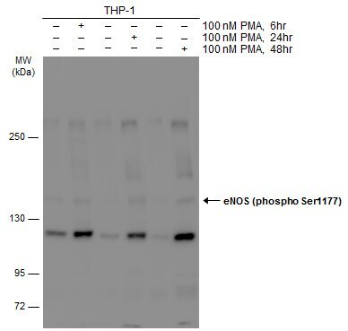
- Western Blot analysis of Phospho-eNOS (Ser1177) was performed by separating 30 µg of various whole cell extracts by 5% SDS-PAGE. Proteins were transferred to a membrane and probed with a Phospho-eNOS (Ser1177) Polyclonal Antibody (Product # PA5-35879) at a dilution of 1:500 and a HRP-conjugated anti-rabbit IgG secondary antibody.
Supportive validation
- Submitted by
- Invitrogen Antibodies (provider)
- Main image
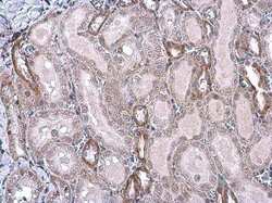
- Experimental details
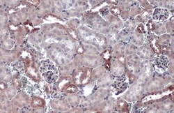
- Immunohistochemistry (Paraffin) analysis of Phospho-eNOS (Ser1177) was performed in paraffin-embedded rat kidney tissue using Phospho-eNOS (Ser1177) Polyclonal Antibody (Product # PA5-35879) at a dilution of 1:500.
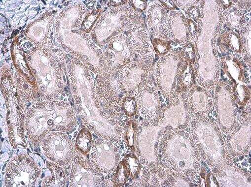
- Submitted by
- Invitrogen Antibodies (provider)
- Main image
- Experimental details
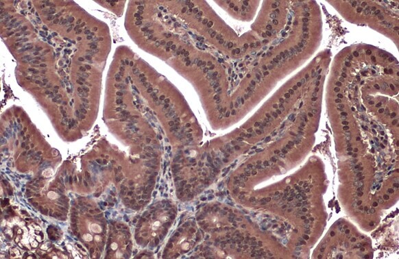
- Immunohistochemistry (Paraffin) analysis of Phospho-eNOS (Ser1177) was performed in paraffin-embedded mouse kidney tissue using Phospho-eNOS (Ser1177) Polyclonal Antibody (Product # PA5-35879) at a dilution of 1:500. Antigen Retrieval: Citrate buffer, pH 6.0, 15 min.

- Submitted by
- Invitrogen Antibodies (provider)
- Main image

- Experimental details
- Immunohistochemistry (Paraffin) analysis of Phospho-eNOS (Ser1177) was performed in paraffin-embedded mouse duodenum tissue using Phospho-eNOS (Ser1177) Polyclonal Antibody (Product # PA5-35879) at a dilution of 1:500. Antigen Retrieval: Citrate buffer, pH 6.0, 15 min.
Supportive validation
- Submitted by
- Invitrogen Antibodies (provider)
- Main image
- Experimental details
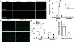
- Fig. 8. ( A and B ) Shear stress-mediated TRPV4-eNOS signaling is diminished in glaucomatous TM cells. ( A ) Normal and glaucomatous primary human TM cells were subjected to different shear stress conditions (0, 1, and 3 dyne/cm 2 ) and treated with NO-binding DAF-FM dye to determine shear stress-mediated NO production. TRPV4 antagonist GSK219 (100 nM) was used to determine the role of TRPV4 in shear stress transduction. High shear stress (3 dyne/cm 2 ) led to increased DAF-FM fluorescence intensity, which was reduced by TRPV4 antagonist GSK219. n = 3 normal and 2 glaucoma. (Scale bar, 50 muM.) ( B ) Mean DAF-FM fluorescence intensity/mum 2 in normal and glaucomatous primary TM cells. *** P < 0.001, n = 3 donor strains per group; one-way ANOVA followed by Bonferroni''s post hoc test. ( C - E ) TRPV4-eNOS coupling is impaired in glaucomatous TM cells. ( C ) Representative image comparing TRPV4-mediated NO production in normal and glaucomatous primary human TM cells using DAF-FM assay. Normal and glaucomatous primary TM cells were pretreated with NO-binding DAF-FM fluorescent dye (green) and then treated with 0.001% DMSO vehicle, 20 nM GSK101, 20 nm GSK101 + 100 muM l -NAME, or 100 muM DETA/NO. n = 3/group (scale bar, 50 mum). ( D ) Quantification of DAF-FM fluorescence intensity/mum 2 in normal and glaucomatous primary TM cells. * P < 0.05, ** P < 0.01 versus the vehicle-treated group, n = 3 cell strains/group; one-way ANOVA followed by Bonferroni''s post hoc test. ( E ) Densi
- Submitted by
- Invitrogen Antibodies (provider)
- Main image
- Experimental details
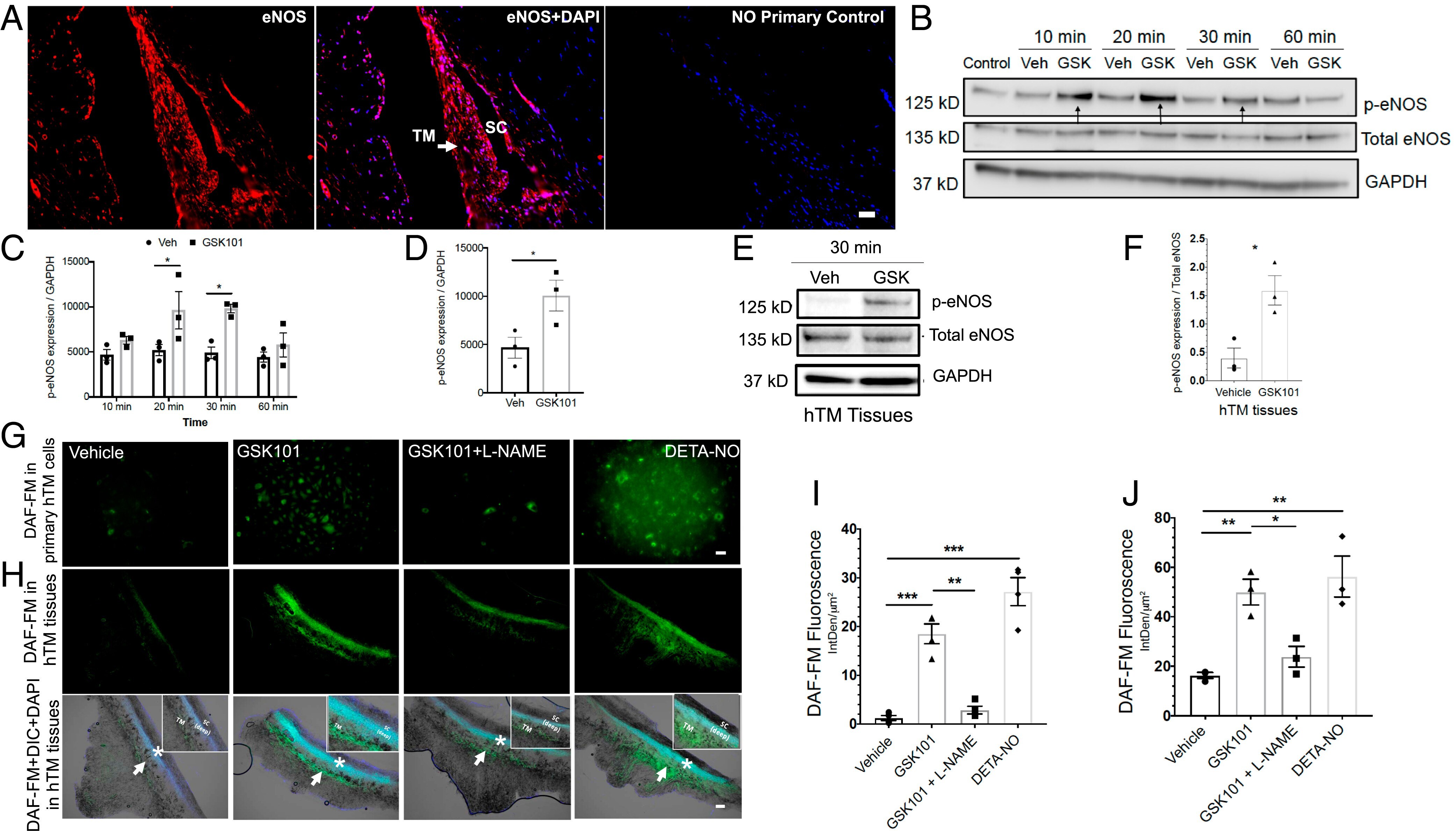
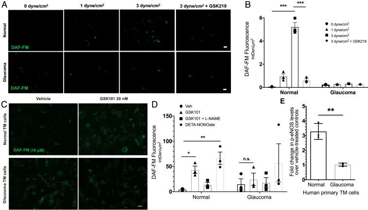
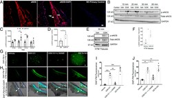
- Fig. 4. TRPV4 channels are functionally coupled to eNOS in human TM cells and tissues. ( A ) Immunohistochemical images showing expression of eNOS (red) in the human TM and SC endothelium ( n = 6). Nuclei were counterstained with DAPI (blue; center image). No primary antibody control ( Right ). (Scale bar, 50 mm.) ( B and C ) TRPV4 activation leads to phosphorylation of eNOS in TM cells. Representative Western blot image ( B ) and densitometric analysis ( C ) showing expression of p-eNOS, total eNOS, and glyceraldehyde 3-phosphate dehydrogenase (GAPDH) in lysates from cultured transformed TM cells (GTM3) treated with 20 nM GSK101 or 0.001% DMSO vehicle for 10, 20, 30, and 60 min intervals. * P < 0.05 versus vehicle at same time point, n = 3/group; unpaired two-tailed t test. ( D ) Densitometric analysis of Western blot for p-eNOS levels in primary human TM cells treated with 20 nM GSK101 or 0.001% DMSO vehicle. * P < 0.05 versus vehicle, n = 3 donors/group; unpaired two-tailed t test. ( E and F ) TRPV4-mediated phosphorylation of eNOS in human donor TM tissues. Representative Western blot ( E ) showing expression of p-eNOS, total eNOS, and GAPDH in ex vivo cultured human TM tissues treated with 20 nM GSK101 and 0.01% DMSO vehicle. Densitometric analysis ( F ) compares levels of p-eNOS over eNOS between the groups. * P < 0.05 versus vehicle, n = 3/group; unpaired two-tailed t test. ( G - J ) TRPV4 activation leads to NO production in primary TM cells and ex vivo cultured human