Explore
 Validate
 Learn
Learn Western blot
 ELISA
ELISA Immunohistochemistry
Antibody data
- Antibody Data
- Antigen structure
- References [0]
- Comments [0]
- Validations
- Western blot [1]
- Immunohistochemistry [2]
Submit
Validation data
Reference
Comment
Report error
- Product number
- LS-B425 - Provider product page

- Provider
- LSBio
- Proper citation
- LifeSpan Cat#LS-B425, RRID:AB_2286611
- Product name
- PathPlus™ SPP1 / Osteopontin Antibody LS-B425
- Antibody type
- Polyclonal
- Description
- Antiserum
- Reactivity
- Human, Mouse, Rat, Canine, Porcine
- Host
- Rabbit
- Storage
- Short term: store at 4°C. Long term: store at -20°C. Avoid freeze-thaw cycles.
No comments: Submit comment
Enhanced validation
- Submitted by
- LSBio (provider)
- Enhanced method
- Genetic validation
- Main image

- Experimental details
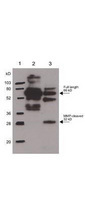
- Anti-Osteopontin Antibody - Western Blot. Rabbit anti-Osteopontin was used at a 1:1000 dilution to detect Osteopontin by western blot. In lane 2, reactivity is shown against 250 ng of human osteopontin. Lane 3 shows reactivity of MMP-cleaved osteopontin. Lane 1 shows the position of molecular weight markers. Use a 1:10000 dilution of HRP conjugated Gt-a-Rabbit IgG (LS-C60865) for detection.
Supportive validation
- Submitted by
- LSBio (provider)
- Enhanced method
- Genetic validation
- Main image

- Experimental details
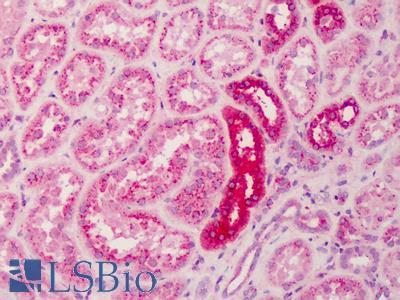
- Anti-Osteopontin antibody IHC of human kidney. Immunohistochemistry of formalin-fixed, paraffin-embedded tissue after heat-induced antigen retrieval. Antibody dilution 1:500.
- Submitted by
- LSBio (provider)
- Enhanced method
- Genetic validation
- Main image

- Experimental details
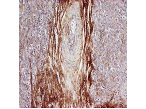
- Rabbit anti-Osteopontin was used at a 1:100-1:300 dilution to detect osteopontin by immunohistochemistry. Osteopontin is a normal component of elastic fibers in the breast (shown heavily stained in this section of human breast tumor cells). There is also weak staining of the extracellular matrix. Osteopontin is not expressed in breast tumor cells, and there is no staining of the breast cells in this section. No antigen retrieval is required.