 Explore
 Validate
 Learn
Learn Western blot
Antibody data
- Antibody Data
- Antigen structure
- References [4]
- Comments [0]
- Validations
- Western blot [2]
Submit
Validation data
Reference
Comment
Report error
- Product number
- MAB14331-100 - Provider product page

- Provider
- R&D Systems
- Product name
- Human Osteopontin/OPN Antibody
- Antibody type
- Monoclonal
- Description
- Protein A or G purified from hybridoma culture supernatant. Detects human Osteopontin in direct ELISAs and Western blots. In direct ELISAs and Western blots, this antibody does not cross-react with recombinant mouse Osteopontin.
- Reactivity
- Human
- Host
- Mouse
- Conjugate
- Unconjugated
- Antigen sequence
NP_000573.1- Isotype
- IgG
- Antibody clone number
- 223126
- Vial size
- 100 ug
- Concentration
- LYOPH
- Storage
- Use a manual defrost freezer and avoid repeated freeze-thaw cycles. 12 months from date of receipt, -20 to -70 °C as supplied. 1 month, 2 to 8 °C under sterile conditions after reconstitution. 6 months, -20 to -70 °C under sterile conditions after reconstitution.
Submitted references Correlation of OPN gene expression with proliferation and apoptosis of ovarian cancer cells and prognosis of patients.
Thrombin-cleaved fragments of osteopontin are overexpressed in malignant glial tumors and provide a molecular niche with survival advantage.
Regional immunity in melanoma: immunosuppressive changes precede nodal metastasis.
Osteopontin induces AP-1-mediated secretion of urokinase-type plasminogen activator through c-Src-dependent epidermal growth factor receptor transactivation in breast cancer cells.
Hu H, Liu Z, Liu C
Oncology letters 2019 Mar;17(3):2788-2794
Oncology letters 2019 Mar;17(3):2788-2794
Thrombin-cleaved fragments of osteopontin are overexpressed in malignant glial tumors and provide a molecular niche with survival advantage.
Yamaguchi Y, Shao Z, Sharif S, Du XY, Myles T, Merchant M, Harsh G, Glantz M, Recht L, Morser J, Leung LL
The Journal of biological chemistry 2013 Feb 1;288(5):3097-111
The Journal of biological chemistry 2013 Feb 1;288(5):3097-111
Regional immunity in melanoma: immunosuppressive changes precede nodal metastasis.
Mansfield AS, Holtan SG, Grotz TE, Allred JB, Jakub JW, Erickson LA, Markovic SN
Modern pathology : an official journal of the United States and Canadian Academy of Pathology, Inc 2011 Apr;24(4):487-94
Modern pathology : an official journal of the United States and Canadian Academy of Pathology, Inc 2011 Apr;24(4):487-94
Osteopontin induces AP-1-mediated secretion of urokinase-type plasminogen activator through c-Src-dependent epidermal growth factor receptor transactivation in breast cancer cells.
Das R, Mahabeleshwar GH, Kundu GC
The Journal of biological chemistry 2004 Mar 19;279(12):11051-64
The Journal of biological chemistry 2004 Mar 19;279(12):11051-64
No comments: Submit comment
Supportive validation
- Submitted by
- R&D Systems (provider)
- Main image
- Experimental details
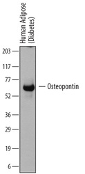
- Detection of Human Osteopontin/OPN by Western Blot. Western blot shows lysates of human adipose (diabetes) tissue. PVDF membrane was probed with 2 µg/mL of Mouse Anti-Human Osteopontin/OPN Monoclonal Antibody (Catalog # MAB14331) followed by HRP-conjugated Anti-Mouse IgG Secondary Antibody (Catalog # HAF007). A specific band was detected for Osteopontin/OPN at approximately 60 to 65 kDa (as indicated). This experiment was conducted under reducing conditions and using Immunoblot Buffer Group 1.

- Submitted by
- R&D Systems (provider)
- Main image

- Experimental details
- Detection of Human Osteopontin/OPN by Simple WesternTM. Simple Western lane view shows lysates of human adipose tissue and human adipose (diabetes) tissue, loaded at 0.2 mg/mL. A specific band was detected for Osteopontin/OPN at approximately 63 kDa (as indicated) using 100 µg/mL of Mouse Anti-Human Osteopontin/OPN Monoclonal Antibody (Catalog # MAB14331). This experiment was conducted under reducing conditions and using the 12-230 kDa separation system. *Non-specific interaction with the 230 kDa Simple Western standard may be seen with this antibody.
