 Explore
 Validate
 Learn
Learn Western blot
Antibody data
- Antibody Data
- Antigen structure
- References [1]
- Comments [0]
- Validations
- Western blot [1]
- Immunocytochemistry [1]
- Immunohistochemistry [1]
Submit
Validation data
Reference
Comment
Report error
- Product number
- MAB808-100 - Provider product page

- Provider
- R&D Systems
- Product name
- Mouse Osteopontin/OPN Antibody
- Antibody type
- Monoclonal
- Description
- Protein A or G purified from cell culture supernatant. Detects mouse Osteopontin/OPN in direct ELISAs.
- Reactivity
- Mouse
- Host
- Rabbit
- Conjugate
- Unconjugated
- Antigen sequence
Q547B5- Isotype
- IgG
- Antibody clone number
- 2139B
- Vial size
- 100 ug
- Storage
- Use a manual defrost freezer and avoid repeated freeze-thaw cycles. 12 months from date of receipt, -20 to -70 °C as supplied. 1 month, 2 to 8 °C under sterile conditions after reconstitution. 6 months, -20 to -70 °C under sterile conditions after reconstitution.
Submitted references Long-term neuronal survival, regeneration, and transient target reconnection after optic nerve crush and mesenchymal stem cell transplantation.
Mesentier-Louro LA, Teixeira-Pinheiro LC, Gubert F, Vasques JF, Silva-Junior AJ, Chimeli-Ormonde L, Nascimento-Dos-Santos G, Mendez-Otero R, Santiago MF
Stem cell research & therapy 2019 Apr 17;10(1):121
Stem cell research & therapy 2019 Apr 17;10(1):121
No comments: Submit comment
Supportive validation
- Submitted by
- R&D Systems (provider)
- Main image

- Experimental details
- Detection of Mouse Osteopontin/OPN by Western Blot. Western blot shows conditioned media from Neuro-2A mouse neuroblastoma cell line (negative control) and L-929 mouse fibroblast cell line. PVDF membrane was probed with 1 µg/mL of Rabbit Anti-Mouse Osteopontin/OPN Monoclonal Antibody (Catalog # MAB808) followed by HRP-conjugated Anti-Rabbit IgG Secondary Antibody (Catalog # HAF008). A specific band was detected for Osteopontin/OPN at approximately 50 kDa (as indicated). This experiment was conducted under reducing conditions and using Immunoblot Buffer Group 1.

Supportive validation
- Submitted by
- R&D Systems (provider)
- Main image

- Experimental details
- Osteopontin/OPN in RAW 264.7 Mouse Cell Line. Osteopontin/OPN was detected in immersion fixed RAW 264.7 mouse monocyte/macrophage cell line using Rabbit Anti-Mouse Osteopontin/OPN Monoclonal Antibody (Catalog # MAB808) at 3 µg/mL for 3 hours at room temperature. Cells were stained using the NorthernLights™ 557-conjugated Anti-Rabbit IgG Secondary Antibody (red; Catalog # NL004) and counterstained with DAPI (blue). Specific staining was localized to cytoplasm. View our protocol for Fluorescent ICC Staining of Non-adherent Cells.

Supportive validation
- Submitted by
- R&D Systems (provider)
- Main image
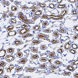
- Experimental details
- Osteopontin/OPN in Mouse Kidney. Osteopontin/OPN was detected in perfusion fixed frozen sections of mouse kidney using Rabbit Anti-Mouse Osteopontin/OPN Monoclonal Antibody (Catalog # MAB808) at 3 µg/mL for 1 hour at room temperature followed by incubation with the Anti-Rabbit IgG VisUCyte™ HRP Polymer Antibody (Catalog # VC003). Tissue was stained using DAB (brown) and counterstained with hematoxylin (blue). Specific staining was localized to convoluted tubules. View our protocol for IHC Staining with VisUCyte HRP Polymer Detection Reagents.
