Explore
 Validate
 Learn
Learn Western blot
Antibody data
- Antibody Data
- Antigen structure
- References [1]
- Comments [0]
- Validations
- Western blot [1]
- Immunohistochemistry [2]
- Flow cytometry [2]
- Other assay [2]
Submit
Validation data
Reference
Comment
Report error
- Product number
- PA5-13494 - Provider product page

- Provider
- Invitrogen Antibodies
- Product name
- Osteopontin Polyclonal Antibody
- Antibody type
- Polyclonal
- Antigen
- Synthetic peptide
- Reactivity
- Human
- Host
- Rabbit
- Isotype
- IgG
- Vial size
- 400 μL
- Concentration
- 2 mg/mL
- Storage
- Store at 4°C short term. For long term storage, store at -20°C, avoiding freeze/thaw cycles.
Submitted references Evaluation of SPP1/osteopontin expression as predictor of recurrence in tamoxifen treated breast cancer.
Göthlin Eremo A, Lagergren K, Othman L, Montgomery S, Andersson G, Tina E
Scientific reports 2020 Jan 29;10(1):1451
Scientific reports 2020 Jan 29;10(1):1451
No comments: Submit comment
Supportive validation
- Submitted by
- Invitrogen Antibodies (provider)
- Main image

- Experimental details
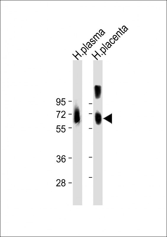
- Western blot analysis of Osteopontin in various lysates. Samples were incubated with Osteopontin polyclonal antibody (Product # PA5-13494) using a dilution of 1:2,000 followed by Goat Anti-Rabbit IgG, (H+L), Peroxidase conjugated at a dilution of 1:10,000. Lysates/proteins: 20 µg per lane. Lane 1: Human plasma lysate; Lane 2: Human placenta lysate. Predicted band size: 35 kDa. Blocking/Dilution buffer: 5% NFDM/TBST.
Supportive validation
- Submitted by
- Invitrogen Antibodies (provider)
- Main image

- Experimental details
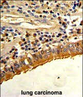
- Immunohistochemistry analysis of Osteopontin in formalin-fixed and paraffin-embedded human lung carcinoma. Samples were incubated with Osteopontin polyclonal antibody (Product # PA5-13494) which was peroxidase-conjugated to the secondary antibody, followed by DAB staining. This data demonstrates the use of this antibody for immunohistochemistry; clinical relevance has not been evaluated.
- Submitted by
- Invitrogen Antibodies (provider)
- Main image
- Experimental details
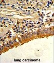
- Immunohistochemistry analysis of Osteopontin in formalin-fixed and paraffin-embedded human lung carcinoma. Samples were incubated with Osteopontin polyclonal antibody (Product # PA5-13494) which was peroxidase-conjugated to the secondary antibody, followed by DAB staining. This data demonstrates the use of this antibody for immunohistochemistry; clinical relevance has not been evaluated.

Supportive validation
- Submitted by
- Invitrogen Antibodies (provider)
- Main image

- Experimental details
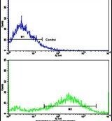
- Flow cytometry analysis of NCI-H292 cells using a SPP1 polyclonal antibody (Product # PA5-13494) (bottom), compared to a negative control cell (top) at a dilution of 1:10-50, followed by a FITC-conjugated goat anti-rabbit antibody
- Submitted by
- Invitrogen Antibodies (provider)
- Main image

- Experimental details
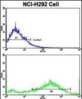
- Flow cytometry of Osteopontin in NCI-H292 cells (bottom histogram). Samples were incubated with Osteopontin polyclonal antibody (Product # PA5-13494) followed by FITC-conjugated goat-anti-rabbit secondary antibody. Negative control cell (top histogram).
Supportive validation
- Submitted by
- Invitrogen Antibodies (provider)
- Main image
- Experimental details
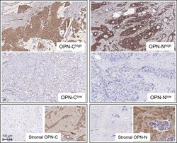
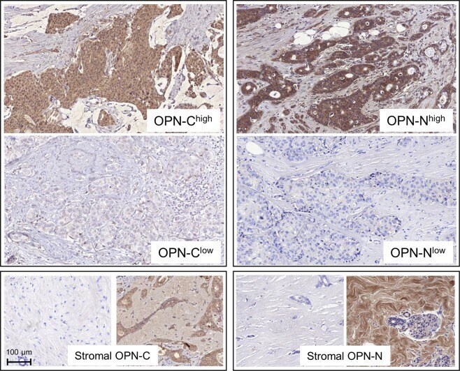
- Figure 3 Micrographs of IHC staining using antibodies against OPN C and N-terminals. All micrographs are snapshots taken with CaseViewer (3DHistech) in x20 magnification. The bottom left scale bar applies to all micrographs.
- Submitted by
- Invitrogen Antibodies (provider)
- Main image

- Experimental details
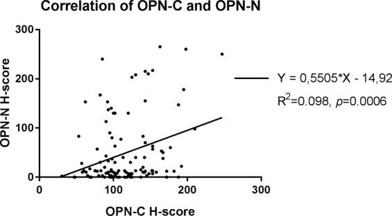
- Figure 5 Correlation between staining results (H-score) of OPN-C and OPN-N expression in tumours. The scatter plot illustrates values for each individual tumour (n = 116).