Explore
 Validate
 Learn
Learn Western blot
 Immunohistochemistry
Antibody data
- Antibody Data
- Antigen structure
- References [0]
- Comments [0]
- Validations
- Immunohistochemistry [1]
Submit
Validation data
Reference
Comment
Report error
- Product number
- NBP2-46876 - Provider product page

- Provider
- Novus Biologicals
- Product name
- Rabbit Polyclonal Collagen II Antibody
- Antibody type
- Polyclonal
- Description
- Immunogen affinity purified. Typically less than 1% cross reactivity against other types of collagens was detected by ELISA against purified standards. Some class specific anti-collagens may be specific for three-dimensional epitopes which may result in diminished reactivity with de
- Reactivity
- Human, Mouse, Rat, Bovine, Sheep
- Host
- Rabbit
- Conjugate
- Green dye
- Isotype
- IgG
- Vial size
- 0.05 mg
- Concentration
- LYOPH
- Storage
- Store lyophilized antibody at 4C. Aliquot reconstituted liquid and store at -20C. Avoid freeze-thaw cycles.
No comments: Submit comment
Supportive validation
- Submitted by
- Novus Biologicals (provider)
- Main image
- Experimental details
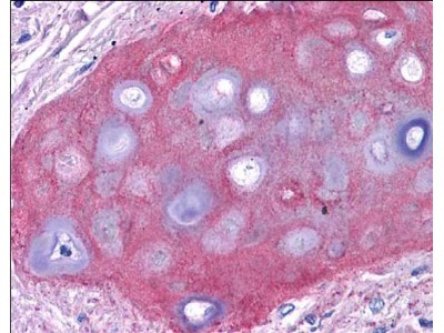
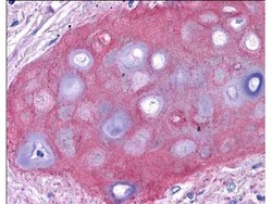
- Immunohistochemistry: Collagen II Antibody [Fluorescein] [NBP2-46876] - Analysis of Tissue: human bronchiolar cartilage (shown). Though not shown, faint to moderate staining of tonsillar squamous epithelium, prostatic stroma, breast, colon, placenta, and dermal connective tissues was also observed. All other tissues, including brain, breast epithelium, colon epithelium, heart, intestine, kidney, liver, lung, skeletal muscle, pancreas, spleen, testis, thymus, thyroid, and uterus were negative for staining. Fixation: formalin fixed paraffin embedded. Antigen retrieval: 0.01 M sodium citrate buffer, pH 6.0 at 99-100C - 20 minutes. Primary antibody: collagen II antibody at 10 ug/mL for 1 h at RT. Secondary antibody: Peroxidase rabbit secondary antibody at 1:10,000 for 45 min at RT. Localization: Collagen II is extracellular. Staining: Collagen II as precipitated red signal with hematoxylin purple nuclear counterstain.