 Explore
 Validate
 Learn
Learn Western blot
 ELISA
ELISA Immunocytochemistry
Antibody data
- Antibody Data
- Antigen structure
- References [0]
- Comments [0]
- Validations
- Immunocytochemistry [1]
- Immunoprecipitation [1]
- Immunohistochemistry [3]
Submit
Validation data
Reference
Comment
Report error
- Product number
- MA5-35215 - Provider product page

- Provider
- Invitrogen Antibodies
- Product name
- CDX2 Recombinant Rabbit Monoclonal Antibody (9E0K5)
- Antibody type
- Monoclonal
- Antigen
- Synthetic peptide
- Description
- Immunogen sequence: MYVSYLLDKD VSMYPSSVRH SGGLNLAPQN FVSPPQYPDY GGYHVAAAAA AAANLDSAQS PGPSWPAAYG APLREDWNGY APGGAAAAAN AVAHGLNGGS
- Reactivity
- Human, Mouse, Rat
- Host
- Rabbit
- Isotype
- IgG
- Antibody clone number
- 9E0K5
- Vial size
- 100 μL
- Concentration
- 1 mg/mL
- Storage
- -20°C, Avoid Freeze/Thaw Cycles
No comments: Submit comment
Supportive validation
- Submitted by
- Invitrogen Antibodies (provider)
- Main image
- Experimental details
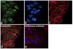
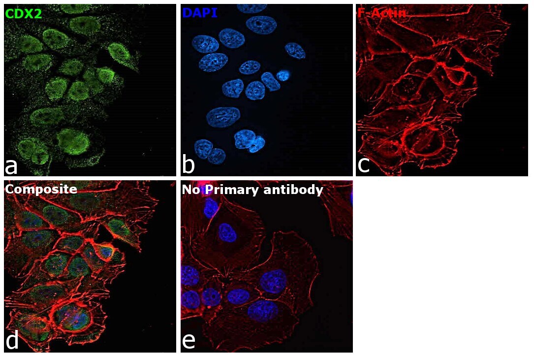
- Immunofluorescence analysis of CDX2 was performed using 70% confluent log phase LoVo cells. The cells were fixed with 4% paraformaldehyde for 10 minutes, permeabilized with 0.1% Triton™ X-100 for 15 minutes, and blocked with 2% BSA for 45 minutes at room temperature. The cells were labeled with CDX2 Recombinant Rabbit Monoclonal Antibody (ARC0450) (Product # MA5-35215) at 5 µg/mL in 0.1% BSA, incubated at 4 degree celsius overnight and then labeled with Donkey anti-Rabbit IgG (H+L) Highly Cross-Adsorbed Secondary Antibody, Alexa Fluor Plus 488 (Product # A32790, 1:2000 dilution), for 45 minutes at room temperature (Panel a: Green). Nuclei (Panel b:Blue) were stained with ProLong™ Diamond Antifade Mountant with DAPI (Product # P36962). F-actin (Panel c: Red) was stained with Rhodamine Phalloidin (Product # R415, 1:300 dilution). Panel d represents the merged image showing nuclear as well as cytoplasmic localization. Panel e represents control cells with no primary antibody to assess background. The images were captured at 60X magnification.
Supportive validation
- Submitted by
- Invitrogen Antibodies (provider)
- Main image
- Experimental details
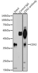
- Immunoprecipitation of CDX2 in 600 μg extracts of 293T cells. Samples were precipitated with 3 μg CDX2 Monoclonal antibody (Product # MA5-35215). Western blot was performed from the immunoprecipitate using CDX2 Monoclonal antibody (Product # MA5-35215) at a dilution of 1:500.

Supportive validation
- Submitted by
- Invitrogen Antibodies (provider)
- Main image

- Experimental details
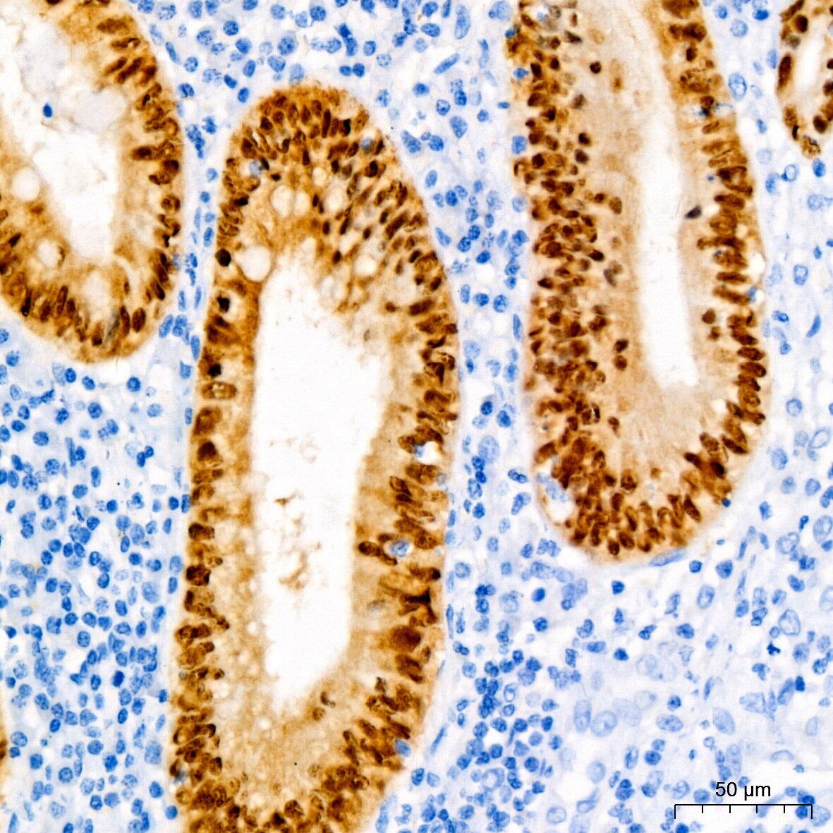
- Immunohistochemistry analysis of CDX2 in paraffin-embedded human appendix. Samples were incubated with CDX2 Monoclonal antibody (Product # MA5-35215) using a dilution of 1:100 (40x lens). Perform high pressure antigen retrieval with 10 mM Tris/EDTA buffer pH 9.0 before commencing with IHC staining protocol.
- Submitted by
- Invitrogen Antibodies (provider)
- Main image
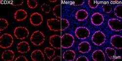
- Experimental details
- Confocal imaging (Immunohistochemistry) of CDX2 in human colon. Samples were incubated with CDX2 Monoclonal antibody (Product # MA5-35215) using a dilution of 1:100 (Red). DAPI was used for nuclear staining (blue). Objective: 40x.

- Submitted by
- Invitrogen Antibodies (provider)
- Main image
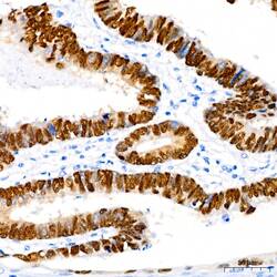
- Experimental details
- Immunohistochemistry analysis of CDX2 in paraffin-embedded human colon carcinoma. Samples were incubated with CDX2 Monoclonal antibody (Product # MA5-35215) using a dilution of 1:100 (40x lens). Perform high pressure antigen retrieval with 10 mM Tris/EDTA buffer pH 9.0 before commencing with IHC staining protocol.
