 Explore
 Validate
 Learn
Learn Western blot
Antibody data
- Antibody Data
- Antigen structure
- References [0]
- Comments [0]
- Validations
- Western blot [1]
- Immunocytochemistry [1]
- Immunohistochemistry [1]
- Flow cytometry [2]
Submit
Validation data
Reference
Comment
Report error
- Product number
- 710395 - Provider product page

- Provider
- Invitrogen Antibodies
- Product name
- Phospho-TSC2 (Ser939) Recombinant Polyclonal Antibody (23HCLC)
- Antibody type
- Polyclonal
- Antigen
- Other
- Reactivity
- Human, Mouse
- Host
- Rabbit
- Isotype
- IgG
- Antibody clone number
- 23HCLC
- Vial size
- 100 µg
- Concentration
- 0.5 mg/mL
- Storage
- Store at 4°C short term. For long term storage, store at -20°C, avoiding freeze/thaw cycles.
No comments: Submit comment
Supportive validation
- Submitted by
- Invitrogen Antibodies (provider)
- Main image
- Experimental details
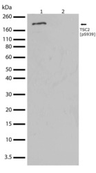
- Western blot analysis of Phospho-Tuberin pSer939 in whole cell extracts from NIH-3T3 cellls treated with Insulin using a Phospho-Tuberin pSer939 Recombinant Rabbit Polyclonal Antibody (Product # 710395) at a dilution of 2 µg/mL. To confirm specificity, competition was performed by preincubation with the phosphopeptide to inhibit antibody binding (lane 2). Samples were detected using chemiluminescence (ECL). Results show a band at ~200kDa.

Supportive validation
- Submitted by
- Invitrogen Antibodies (provider)
- Main image

- Experimental details
- Immunofluorescent analysis of Phospho-Tuberin pSer939 in U2OS cells using a Phospho-Tuberin pSer939 Recombinant Rabbit Polyclonal Antibody (Product # 710395) followed by detection using an Alexa Fluor 488-conjugated Goat anti-Rabbit secondary antibody (green) (Image A). Nuclei were stained using DAPI (blue) (Image D) and actin stained with Alexa Fluor 594 phalloidin (red) (image B). Image C is a composite image showing nuclear localization of Tuberin (pS939).

Supportive validation
- Submitted by
- Invitrogen Antibodies (provider)
- Main image

- Experimental details
- Immunohistochemistry analysis of TSC2 (PS939) showing staining in the cytoplasm of paraffin-embedded human kidney tissue (right) compared to a negative control without primary antibody (left). To expose target proteins, antigen retrieval was performed using 10mM sodium citrate (pH 6.0), microwaved for 8-15 min. Following antigen retrieval, tissues were blocked in 3% H2O2-methanol for 15 min at room temperature, washed with ddH2O and PBS, and then probed with a TSC2 (PS939) Recombinant Rabbit Polyclonal Antibody (Product # 710395) diluted in 3% BSA-PBS at a dilution of 1:100 for 1 hour at 37ºC in a humidified chamber. Tissues were washed extensively in PBST and detection was performed using an HRP-conjugated secondary antibody followed by colorimetric detection using a DAB kit. Tissues were counterstained with hematoxylin and dehydrated with ethanol and xylene to prep for mounting.

Supportive validation
- Submitted by
- Invitrogen Antibodies (provider)
- Main image

- Experimental details
- Fixed and permeabilized HeLa and HepG2 cells were stained with TSC pS939 Recombinant Rabbit Polyclonal Antibody (Product # 710395), followed by Alexa Fluor 488 Goat anti-Rabbit Ig (right peak). Cells stained with TSC pS939 Recombinant Rabbit Polyclonal Antibody after incubation with phosphopeptide (middle peak) and Isotype control (left peak).

- Submitted by
- Invitrogen Antibodies (provider)
- Main image

- Experimental details
- Flow cytometry analysis of Phospho-Tuberin pSer939 in HepG2 cells using a Phospho-Tuberin pSer939 Recombinant Rabbit Polyclonal Antibody (Product # 710395). Cells were fixed and permeabilized using FIX & PERM (Product # GAS-004) reagent, and detection was performed using an Alexa Fluor 488 Goat anti-Rabbit IgG (right peak) compared to an isotype control (left peak).
