Explore
 Validate
 Learn
Learn Western blot
Antibody data
- Antibody Data
- Antigen structure
- References [0]
- Comments [0]
- Validations
- Western blot [3]
- Immunocytochemistry [1]
- Immunohistochemistry [1]
Submit
Validation data
Reference
Comment
Report error
- Product number
- PA5-13364 - Provider product page

- Provider
- Invitrogen Antibodies
- Product name
- TSC2 Polyclonal Antibody
- Antibody type
- Polyclonal
- Antigen
- Synthetic peptide
- Reactivity
- Human
- Host
- Rabbit
- Isotype
- IgG
- Vial size
- 400 µL
- Concentration
- 0.5 mg/mL
- Storage
- -20° C, Avoid Freeze/Thaw Cycles
No comments: Submit comment
Supportive validation
- Submitted by
- Invitrogen Antibodies (provider)
- Main image

- Experimental details
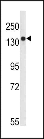
- Western blot analysis using a Tuberin polyclonal antibody (Product # PA5-13364) in Ramos cell lysates (35 µg per lane).
- Submitted by
- Invitrogen Antibodies (provider)
- Main image

- Experimental details
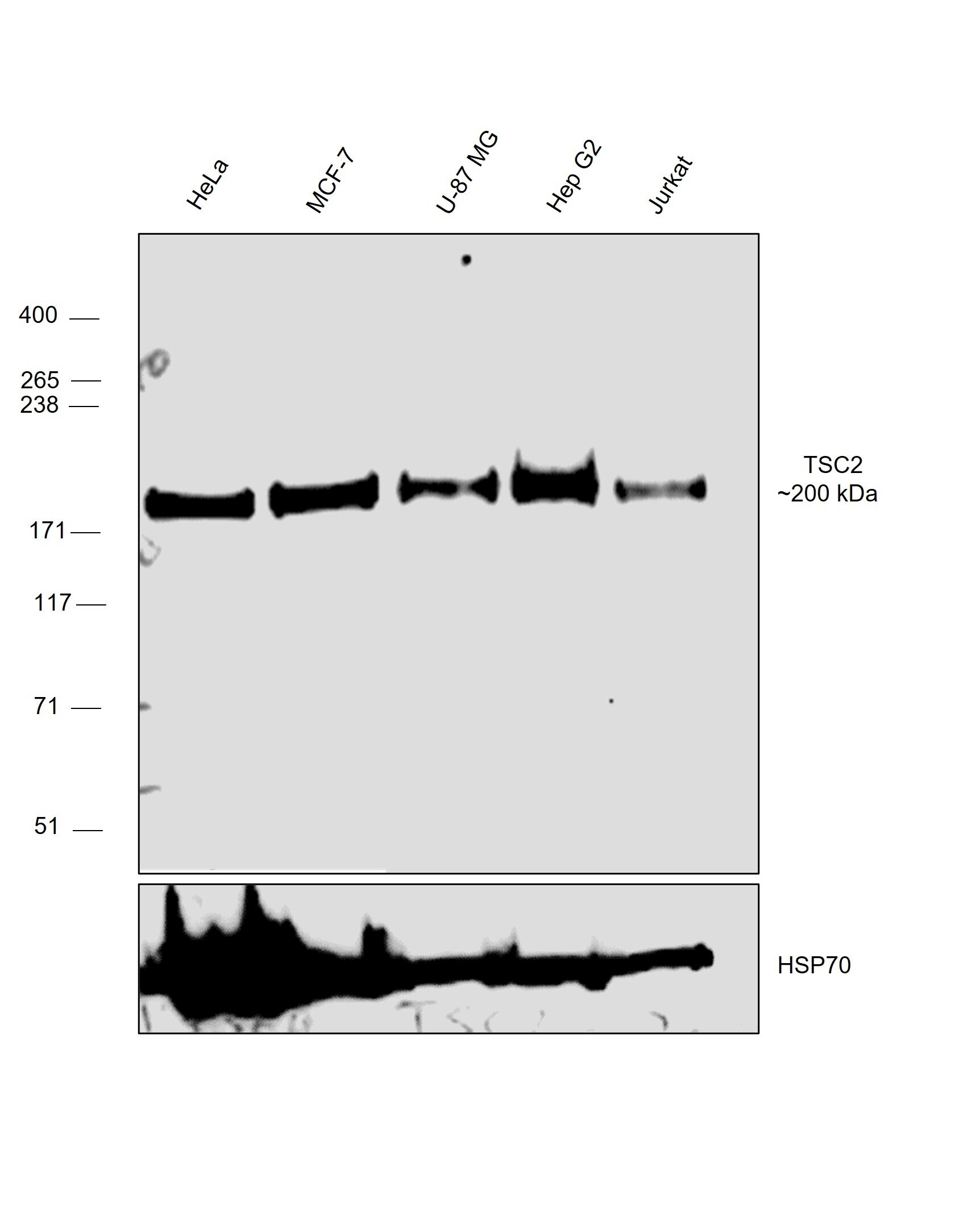
- Western blot was performed using Anti-TSC2 Polyclonal Antibody (Product # PA5-13364) and a 200 kDa band corresponding to TSC2 was observed across the panel tested. Whole cell extracts (30 µg lysate) of HeLa (Lane 1), MCF7 (Lane 2), U-87 MG (Lane 3), Hep G2 (Lane 4), Jurkat (Lane 5) were electrophoresed using NuPAGE™ 3-8% Tris-Acetate Protein Gel (Product # EA0378BOX), 10 well. Resolved proteins were then transferred onto a nitrocellulose membrane (Product # IB23001) by iBlot® 2 Dry Blotting System (Product # IB21001). The blot was probed with the primary antibody (1:1000 dilution) and detected by chemiluminescence with Goat anti-Rabbit IgG (H+L) Superclonal™ Recombinant Secondary Antibody, HRP (Product # A27036, 1:20,000 dilution) using the iBright™ FL1500 Imaging System (Product # A44115). Chemiluminescent detection was performed using SuperSignal™ West Pico PLUS Chemiluminescent Substrate (Product # 34580).
- Submitted by
- Invitrogen Antibodies (provider)
- Main image

- Experimental details
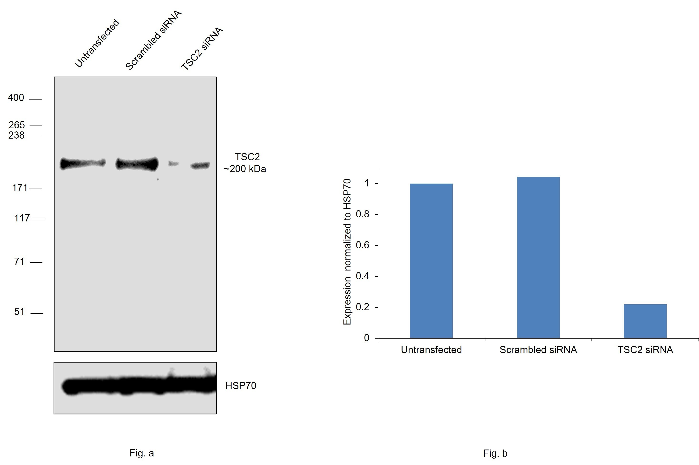
- Knockdown of TSC2 was achieved by transfecting HeLa with TSC2 specific siRNAs (Silencer® select Product # S502596, S502595). Western blot analysis (Fig. a) was performed using Whole cell extracts from the TSC2 knockdown cells (lane 3), non-targeting scrambled siRNA transfected cells (lane 2) and untransfected cells (lane 1). The blot was probed with TSC2 Polyclonal Antibody (Product # PA5-13364, 1:1000 dilution) and Goat anti-Rabbit IgG (H+L) Superclonal™ Recombinant Secondary Antibody, HRP (Product # A27036, 1:20,000 dilution). Densitometric analysis of this western blot is shown in histogram (Fig. b). Decrease in signal upon siRNA mediated knock down confirms that antibody is specific to TSC2.
Supportive validation
- Submitted by
- Invitrogen Antibodies (provider)
- Main image
- Experimental details
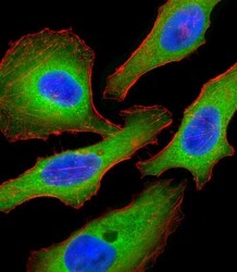
- Immunofluorescent analysis of HeLa cells using a Tuberin polyclonal antibody (Product # PA5-13364). HeLa cells were fixed with 4% PFA (20 min), permeabilized with Triton X-100 (0.1%, 10 min), then incubated with a Tuberin polyclonal antibody (Product # PA5-13364) (1:25, 1 hr at 37°C). Primary antibody was detected with fluor-conjugated donkey anti-rabbit secondary antibody (green) at 1:400 dilution for 50 min at 37°C). Actin filaments have been labeled with dye-conjugated phalloidin (red). Nuclei were counterstained with DAPI (blue) (10 µg/mL, 10 min).

Supportive validation
- Submitted by
- Invitrogen Antibodies (provider)
- Main image
- Experimental details
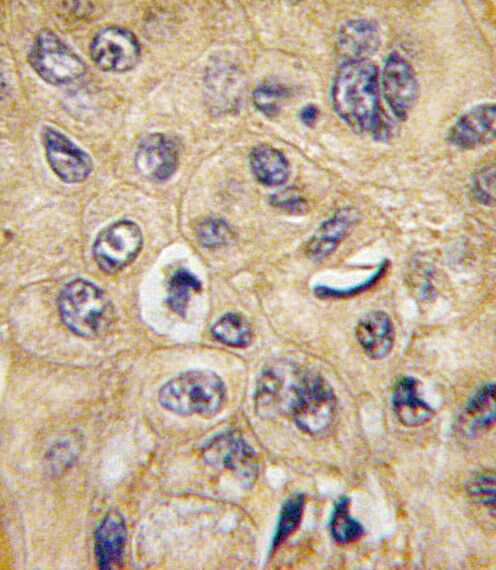
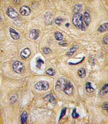
- Immunohistochemical analysis of formalin-fixed, paraffin-embedded human hepatocarcinoma tissue using a Tuberin polyclonal antibody (Product # PA5-13364), followed by HRP-conjugated secondary antibody and DAB staining.