Explore
 Validate
 Learn
Learn Western blot
 Immunocytochemistry
Antibody data
- Antibody Data
- Antigen structure
- References [0]
- Comments [0]
- Validations
- Western blot [1]
- Immunocytochemistry [3]
- Immunohistochemistry [1]
Submit
Validation data
Reference
Comment
Report error
- Product number
- LS-B8056 - Provider product page

- Provider
- LSBio
- Product name
- IHC-plus™ MBP / Myelin Basic Protein Antibody (clone 7D2) LS-B8056
- Antibody type
- Monoclonal
- Description
- Ascites
- Reactivity
- Rat
- Host
- Mouse
- Isotype
- IgG
- Antibody clone number
- 7D2
- Storage
- Short term: store at 4°C. Long term: aliquot and store at -20°C. Avoid freeze-thaw cycles.
No comments: Submit comment
Enhanced validation
- Submitted by
- LSBio (provider)
- Enhanced method
- Genetic validation
- Main image
- Experimental details
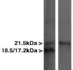
- Myelin Basic Protein Antibody - Blots of crude rat spinal cord homogenate blotted with MBP Antibody (right lane) and another MBP antibody (left lane). The MBP Antibody monoclonal binds only the largest 21.5kDa transcript, while the other MBP antibody binds all three transcripts.

Supportive validation
- Submitted by
- LSBio (provider)
- Enhanced method
- Genetic validation
- Main image

- Experimental details
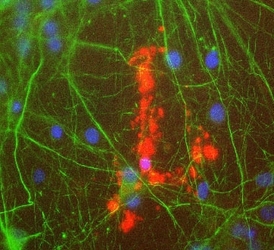
- Rat mixed neuron/glial cultures stained with Myelin Basic Protein antibody (red), and also with chicken antibody to neurofilament NF-L CPCA-NF-L (green). Blue is a DNA stain. Note that the MBP antibody stains an oligodendrocyte and some membrane shed from this cell. Other cells in the field include neurons, astrocytes, microglia and fibroblasts, all of which are completely negative for MBP, though the neuronal processes can be seen with the NF-L antibody.

- Submitted by
- LSBio (provider)
- Main image

- Experimental details
- Rat mixed neuron/glial cultures stained with Myelin Basic Protein antibody (red), and also with chicken antibody to neurofilament NF-L CPCA-NF-L (green). Blue is a DNA stain. Note that the MBP antibody stains an oligodendrocyte and some membrane shed from this cell. Other cells in the field include neurons, astrocytes, microglia and fibroblasts, all of which are completely negative for MBP, though the neuronal processes can be seen with the NF-L antibody.
Supportive validation
- Submitted by
- LSBio (provider)
- Enhanced method
- Genetic validation
- Main image
- Experimental details
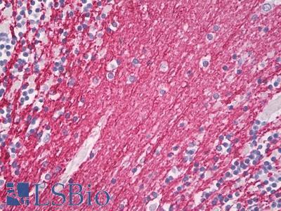
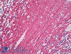
- Anti-Myelin Basic Protein / MBP antibody IHC of human brain, cerebellum, white matter. Immunohistochemistry of formalin-fixed, paraffin-embedded tissue after heat-induced antigen retrieval. Antibody dilution 1:100.