 Explore
 Validate
 Learn
Learn Western blot
 Immunocytochemistry
Antibody data
- Antibody Data
- Antigen structure
- References [1]
- Comments [0]
- Validations
- Western blot [1]
- Immunohistochemistry [1]
Submit
Validation data
Reference
Comment
Report error
- Product number
- NBP1-05204 - Provider product page

- Provider
- Novus Biologicals
- Proper citation
- Novus Cat#NBP1-05204, RRID:AB_1556321
- Product name
- Mouse Monoclonal MBP Antibody
- Antibody type
- Monoclonal
- Description
- Affinity purified. The MBP Antibody (7D2) antibody binds only the 21.5kDa and 18.5kDa rat MBP isotypes, but all four isotypes of human and bovine MBP.
- Reactivity
- Human, Mouse, Rat, Bovine, Porcine
- Host
- Mouse
- Isotype
- IgG
- Vial size
- 0.1 ml
- Concentration
- 1 mg/ml
- Storage
- Store at 4C short term. Aliquot and store at -20C long term. Avoid freeze-thaw cycles.
Submitted references A blend containing docosahexaenoic acid, arachidonic acid, vitamin B12, vitamin B9, iron and sphingomyelin promotes myelination in an in vitro model.
Hauser J, Sultan S, Rytz A, Steiner P, Schneider N
Nutritional neuroscience 2019 Feb 26;:1-15
Nutritional neuroscience 2019 Feb 26;:1-15
No comments: Submit comment
Supportive validation
- Submitted by
- Novus Biologicals (provider)
- Main image
- Experimental details
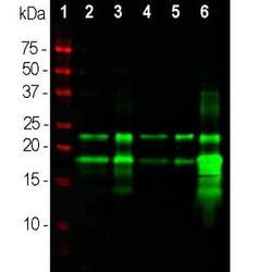
- Western Blot: MBP Antibody (7D2) [NBP1-05204] - Analysis of different tissue lysates using mouse mAb to MBP, NBP1-05204, dilution 1:10,000 in green: [1] protein standard (red), [2] rat brain, [3] rat spinal cord, [4] mouse brain, [5] mouse spinal cord, [6] cow spinal cord. Bands at 21.5kDa and 18.5kDa are the two larger transcripts from the MBP gene, showing that the epitope of this antibody depends on the sequence encoded by exon 2. Note that monoclonal NBP1-05203 binds all four rat MBP transcripts running at 21.5kDa, 18.5kDa, 17kDa and 14kDa, so that this antibody binds to the core sequence of human and rodent MBP.

Supportive validation
- Submitted by
- Novus Biologicals (provider)
- Main image
- Experimental details
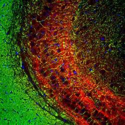
- Immunohistochemistry: MBP Antibody (7D2) [NBP1-05204] - Analysis rat brain hippocampal section stained with mouse mAb to myelin basic protein (MBP), NBP1-05204, dilution 1:5,000 in green, and costained with rabbit pAb to NF-M, RPCA-NF-M, dilution 1:2,000, in red. The MBP antibody stains myelin sheathes around axons, while the NF-M antibody labels dendrites and axons of neuronal cells.
