Explore
 Validate
 Learn
Learn Western blot
Antibody data
- Antibody Data
- Antigen structure
- References [1]
- Comments [0]
- Validations
- Western blot [1]
- Immunocytochemistry [1]
- Immunohistochemistry [1]
- Other assay [1]
Submit
Validation data
Reference
Comment
Report error
- Product number
- MA1-25034 - Provider product page

- Provider
- Invitrogen Antibodies
- Product name
- MBP Monoclonal Antibody (22)
- Antibody type
- Monoclonal
- Antigen
- Other
- Description
- Store product as a concentrated solution. Centrifuge briefly prior to opening the vial.
- Reactivity
- Human, Mouse, Rat
- Host
- Mouse
- Isotype
- IgG
- Antibody clone number
- 22
- Vial size
- 1 mL
- Concentration
- Conc. Not Determined
- Storage
- Store at 4°C short term. For long term storage, store at -20°C, avoiding freeze/thaw cycles.
Submitted references Lasting alterations induced in glial cell phenotypes by short exposure to alcohol during embryonic development in zebrafish.
Chatterjee D, Mahabir S, Chatterjee D, Gerlai R
Addiction biology 2021 Jan;26(1):e12867
Addiction biology 2021 Jan;26(1):e12867
No comments: Submit comment
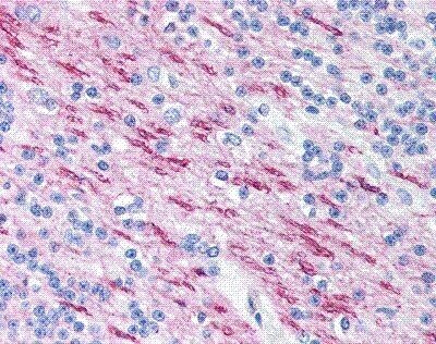
Supportive validation
- Submitted by
- Invitrogen Antibodies (provider)
- Main image
- Experimental details
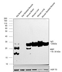
- Western blot was performed using MBP Monoclonal Antibody (22) (Product # MA1-25034) and a 18-30 kDa band corresponding to MBP was observed across tissues tested. Immuno-depleted tissue extracts (30 µg lysate) of Rat Brain (Lane 1), Rat Skeletal Muscle (Lane 2), Mouse Brain (Lane 3), Mouse Liver (Lane 4), Mouse Lung (Lane 5), Mouse Skeletal Muscle (Lane 6) were electrophoresed using NuPAGE™ 4-12% Bis-Tris Protein Gel (Product # NP0322BOX). Resolved proteins were then transferred onto a nitrocellulose membrane (Product # IB33001) by iBlot™ 3 Western Blot Transfer Device (Product # IB31001). The blot was probed with the primary antibody (1:1,000) and detected by chemiluminescence with Goat anti-Mouse IgG (H+L) Superclonal™ Recombinant Secondary Antibody, HRP (Product # A28177, 1:20,000) using the iBright™ FL1500 Imaging System (Product # A44115). Chemiluminescent detection was performed using SuperSignal™ West Pico PLUS Chemiluminescent Substrate (Product # 34580). A 25 kDa band corresponding to IgG light chain was observed in all mouse tissue lysates.
Supportive validation
- Submitted by
- Invitrogen Antibodies (provider)
- Main image

- Experimental details
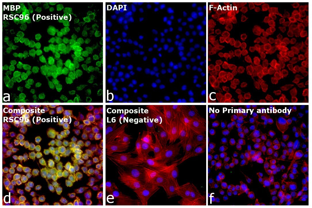
- Immunofluorescence analysis of MBP was performed using 70% confluent log phase RSC96 cells. The cells were fixed with 4% paraformaldehyde for 10 minutes, permeabilized with 0.1% Triton™ X-100 for 15 minutes, and blocked with 2% BSA for 1 hour at room temperature. The cells were labeled with MBP Monoclonal Antibody (22) (Product # MA1-25034, 1:100) in 0.1% BSA, incubated at 4 degree celsius overnight and then labeled with Donkey anti-Mouse IgG (H+L) Highly Cross-Adsorbed Secondary Antibody, Alexa Fluor™ Plus 488 (Product # A32766, 1:2,000), for 45 minutes at room temperature (Panel a: Green). Nuclei (Panel b: Blue) were stained with ProLong™ Diamond Antifade Mountant with DAPI (Product # P36962). F-actin (Panel c: Red) was stained withRhodamine Phalloidin (Product # R415, 1:300). Panel d represents the merged image showing cytoplasmic side of myelin membrane localization. Panel e represents no pick up. Panel f represents control cells with no primary antibody to assess background. The images were captured at 40x magnification.
Supportive validation
- Submitted by
- Invitrogen Antibodies (provider)
- Main image
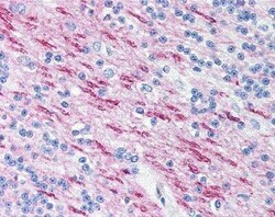
- Experimental details
- Immunohistochemistry (Paraffin) analysis of MBP in white matter in human brain tissue using MBP Monoclonal Antibody (22) (Product # MA1-25034).
Supportive validation
- Submitted by
- Invitrogen Antibodies (provider)
- Main image
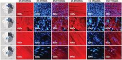
- Experimental details
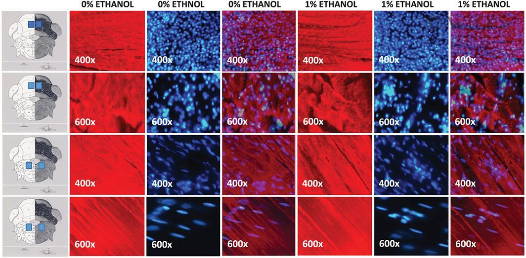
- 4 FIGURE Zebrafish hindbrain stained with anti-MAB antibody. Left most panel shows the schematic diagrams of the corresponding brain areas from where photomicrographs were taken. First column of photomicrographs shows control adult zebrafish hindbrain; second column--DAPI stain applied to the corresponding area; and third column--superimposition of anti-MAB antibody and DAPI stained photomicrographs. Fourth column--adult zebrafish exposed to 1% alcohol (vol/vol) during their embryonic development: hindbrain, stained with anti-MBP antibody; fifth column--DAPI stain applied to the corresponding area; sixth column--superimposition of anti-MAB antibody and DAPI stained photomicrographs. The embryonic alcohol concentration (0 or 1% vol/vol) employed is indicated above the columns of photomicrographs. Magnification of the photomicrographs is also indicated