Explore
 Validate
 Learn
Learn Western blot
Antibody data
- Antibody Data
- Antigen structure
- References [1]
- Comments [0]
- Validations
- Western blot [2]
- Immunocytochemistry [1]
- Immunohistochemistry [3]
Submit
Validation data
Reference
Comment
Report error
- Product number
- MAB42282 - Provider product page

- Provider
- R&D Systems
- Product name
- Human/Mouse/Rat MBP Antibody
- Antibody type
- Monoclonal
- Description
- Protein A or G purified from hybridoma culture supernatant. Detects human, mouse, and rat MBP
- Reactivity
- Human, Mouse, Rat
- Host
- Mouse
- Conjugate
- Unconjugated
- Antigen sequence
P02686- Isotype
- IgG
- Antibody clone number
- 932908
- Vial size
- 100 ug
- Storage
- Use a manual defrost freezer and avoid repeated freeze-thaw cycles. 12 months from date of receipt, -20 to -70 °C as supplied. 1 month, 2 to 8 °C under sterile conditions after reconstitution. 6 months, -20 to -70 °C under sterile conditions after reconstitution.
Submitted references 5-HT1a activation in PO/AH area induces therapeutic hypothermia in a rat model of intracerebral hemorrhage.
Liang T, Chen Q, Li Q, Li R, Tang J, Hu R, Zhong J, Ge H, Liu X, Hua F
Oncotarget 2017 Sep 26;8(43):73613-73626
Oncotarget 2017 Sep 26;8(43):73613-73626
No comments: Submit comment
Supportive validation
- Submitted by
- R&D Systems (provider)
- Main image

- Experimental details
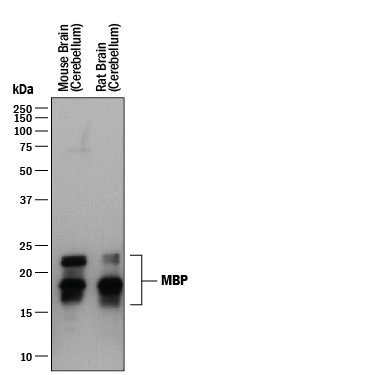
- Detection of Mouse and Rat MBP by Western Blot. Western blot shows lysates of mouse brain (cerebellum) tissue and rat brain (cerebellum) tissue. PVDF membrane was probed with 0.1 µg/mL of Mouse Anti-Human/Mouse/Rat MBP Monoclonal Antibody (Catalog # MAB42282) followed by HRP-conjugated Anti-Mouse IgG Secondary Antibody (Catalog # HAF018). Specific bands were detected for MBP at approximately 15-22 kDa (as indicated). This experiment was conducted under reducing conditions and using Immunoblot Buffer Group 1.
- Submitted by
- R&D Systems (provider)
- Main image
- Experimental details
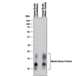
- Detection of Human MBP by Western Blot. Western blot shows lysates of mouse brain (cerebellum) tissue and rat brain (cerebellum). PVDF membrane was probed with 0.1 µg/mL of Mouse Anti-Human/Mouse/Rat MBP Monoclonal Antibody (Catalog # MAB42282) followed by HRP-conjugated Anti-Mouse IgG Secondary Antibody (Catalog # HAF018). Specific bands were detected for MBP at approximately 15-22 kDa (as indicated). This experiment was conducted under reducing conditions and using Immunoblot Buffer Group 1.

Supportive validation
- Submitted by
- R&D Systems (provider)
- Main image

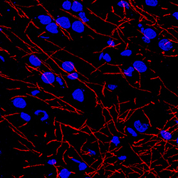
- Experimental details
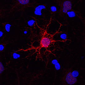
- MBP in Rat Cortical Stem Cells. MBP was detected in immersion fixed rat cortical stem cells differentiated for 7 days to oligodendrocytes using Mouse Anti-Human/Mouse/Rat MBP Monoclonal Antibody (Catalog # MAB42282) at 10 µg/mL for 3 hours at room temperature. Cells were stained using the NorthernLights™ 557-conjugated Anti-Mouse IgG Secondary Antibody (red; Catalog # NL007) and counterstained with DAPI (blue). Specific staining was localized to cell surfaces, cytoplasm, and nuclei. View our protocol for Fluorescent ICC Staining of Stem Cells on Coverslips.
Supportive validation
- Submitted by
- R&D Systems (provider)
- Main image
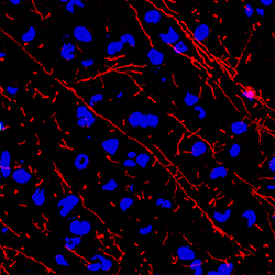
- Experimental details
- MBP in Mouse Brain. MBP was detected in perfusion fixed frozen sections of mouse brain using Mouse Anti-Human/Mouse/Rat MBP Monoclonal Antibody (Catalog # MAB42282) at 0.1 µg/mL overnight at 4 °C. Tissue was stained using the NorthernLights™ 557-conjugated Anti-Mouse IgG Secondary Antibody (red; Catalog # NL007) and counterstained with DAPI (blue). Specific staining was localized to myelinated fibers. View our protocol for Fluorescent IHC Staining of Frozen Tissue Sections.

- Submitted by
- R&D Systems (provider)
- Main image
- Experimental details
- MBP in Rat Brain. MBP was detected in perfusion fixed frozen sections of rat brain using Mouse Anti-Human/Mouse/Rat MBP Monoclonal Antibody (Catalog # MAB42282) at 0.1 µg/mL overnight at 4 °C. Tissue was stained using the NorthernLights™ 557-conjugated Anti-Mouse IgG Secondary Antibody (red; Catalog # NL007) and counterstained with DAPI (blue). Specific staining was localized to myelinated fibers. View our protocol for Fluorescent IHC Staining of Frozen Tissue Sections.

- Submitted by
- R&D Systems (provider)
- Main image

- Experimental details
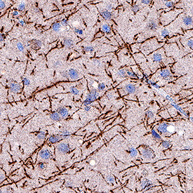
- MBP in Human Brain. MBP was detected in immersion fixed paraffin-embedded sections of human brain (cortex) using Mouse Anti-Human/Mouse/Rat MBP Monoclonal Antibody (Catalog # MAB42282) at 1.7 µg/mL for 1 hour at room temperature followed by incubation with the Anti-Mouse IgG VisUCyte™ HRP Polymer Antibody (Catalog # VC001). Before incubation with the primary antibody, tissue was subjected to heat-induced epitope retrieval using Antigen Retrieval Reagent-Basic (Catalog # CTS013). Tissue was stained using DAB (brown) and counterstained with hematoxylin (blue). Specific staining was localized to neuronal processes. View our protocol for IHC Staining with VisUCyte HRP Polymer Detection Reagents.