 Explore
 Validate
 Learn
Learn Western blot
 Immunohistochemistry
Antibody data
- Antibody Data
- Antigen structure
- References [0]
- Comments [0]
- Validations
- Immunohistochemistry [4]
Submit
Validation data
Reference
Comment
Report error
- Product number
- NBP2-61938 - Provider product page

- Provider
- Novus Biologicals
- Product name
- Rat Monoclonal MBP Antibody
- Antibody type
- Monoclonal
- Description
- Protein G purified.
- Reactivity
- Human, Bovine, Chicken/Avian
- Host
- Rat
- Isotype
- IgG
- Vial size
- 0.1 mg
- Concentration
- 1 mg/ml
- Storage
- Store at 4C short term. Aliquot and store at -20C long term. Avoid freeze-thaw cycles.
No comments: Submit comment
Supportive validation
- Submitted by
- Novus Biologicals (provider)
- Main image

- Experimental details
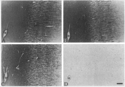
- Immunohistochemistry: MBP Antibody ([MBP14]) - Azide and BSA Free [NBP2-61938] - Clone MBP 14 used to detect myelinated structures in MS plaques by IHC-P. .Clone 14 (B), and clone 2 (C) recognized all myelinated structures in control brains, whereas no immunoreactivity was detected by EP antiserum.
- Submitted by
- Novus Biologicals (provider)
- Main image
- Experimental details
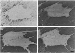
- Immunohistochemistry: MBP Antibody ([MBP14]) - Azide and BSA Free [NBP2-61938] - Clone MBP 14 used to detect myelinated structures in MS plaques by IHC-P. C. Clone 14 recognized all myelinated structures.
- Submitted by
- Novus Biologicals (provider)
- Main image
- Experimental details
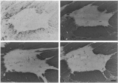
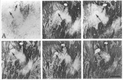
- Immunohistochemistry: MBP Antibody ([MBP14]) - Azide and BSA Free [NBP2-61938] - Clone MBP 14 used to detect myelinated structures in MS plaques by IHC-P. Serial sections of paraffin-embedded MS tissue was immunostained with anti-EP(A), clone26(B), clone2(C), clone14(D), clone12(E), or clone22(F) . Only abnormal myelin tissues were strongly stained by anti-EP, whereas all other antibodies strongly stained the normal myelin surrounding the plaque area.
- Submitted by
- Novus Biologicals (provider)
- Main image
- Experimental details
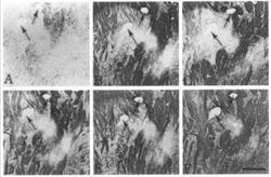
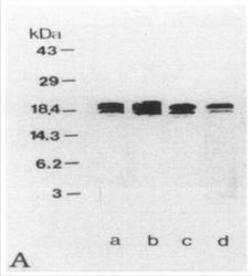
- Immunohistochemistry: MBP Antibody ([MBP14]) - Azide and BSA Free [NBP2-61938] - Western Blot of MBP Clone 14 used in a comparative study between anti-MBP clones and anti-EP, to check the specificity of EP antiserum as examined by Western blot. Lane a, anti-whole hMBP antibody; lane b, clone 14, lane c, clone 2; lane d, EP antiserum. All antibodies detected a major 18.5-kDa band and a weaker 17.2-kDa band in extracts of normal human brain homogenates.
