Explore
 Validate
 Learn
Learn Western blot
 ELISA
ELISAAntibody data
- Antibody Data
- Antigen structure
- References [2]
- Comments [0]
- Validations
- Western blot [3]
- Immunocytochemistry [2]
- Immunohistochemistry [2]
- Flow cytometry [2]
- Other assay [2]
Submit
Validation data
Reference
Comment
Report error
- Product number
- MA5-15922 - Provider product page

- Provider
- Invitrogen Antibodies
- Product name
- MBP Monoclonal Antibody (2H9)
- Antibody type
- Monoclonal
- Antigen
- Purifed from natural sources
- Description
- MA5-15922 targets MBP in indirect ELISA, FACS, IF, IHC, and WB applications and shows reactivity with Human samples. The MA5-15922 immunogen is purified recombinant fragment of human MBP expressed in E. Coli. . MA5-15922 detects MBP which has a predicted molecular weight of approximately 33kDa.
- Reactivity
- Human, Mouse, Rat
- Host
- Mouse
- Isotype
- IgG
- Antibody clone number
- 2H9
- Vial size
- 100 μL
- Concentration
- Conc. not determined
- Storage
- Store at 4°C short term. For long term storage, store at -20°C, avoiding freeze/thaw cycles.
Submitted references Lasmiditan and 5-Hydroxytryptamine in the rat trigeminal system; expression, release and interactions with 5-HT(1) receptors.
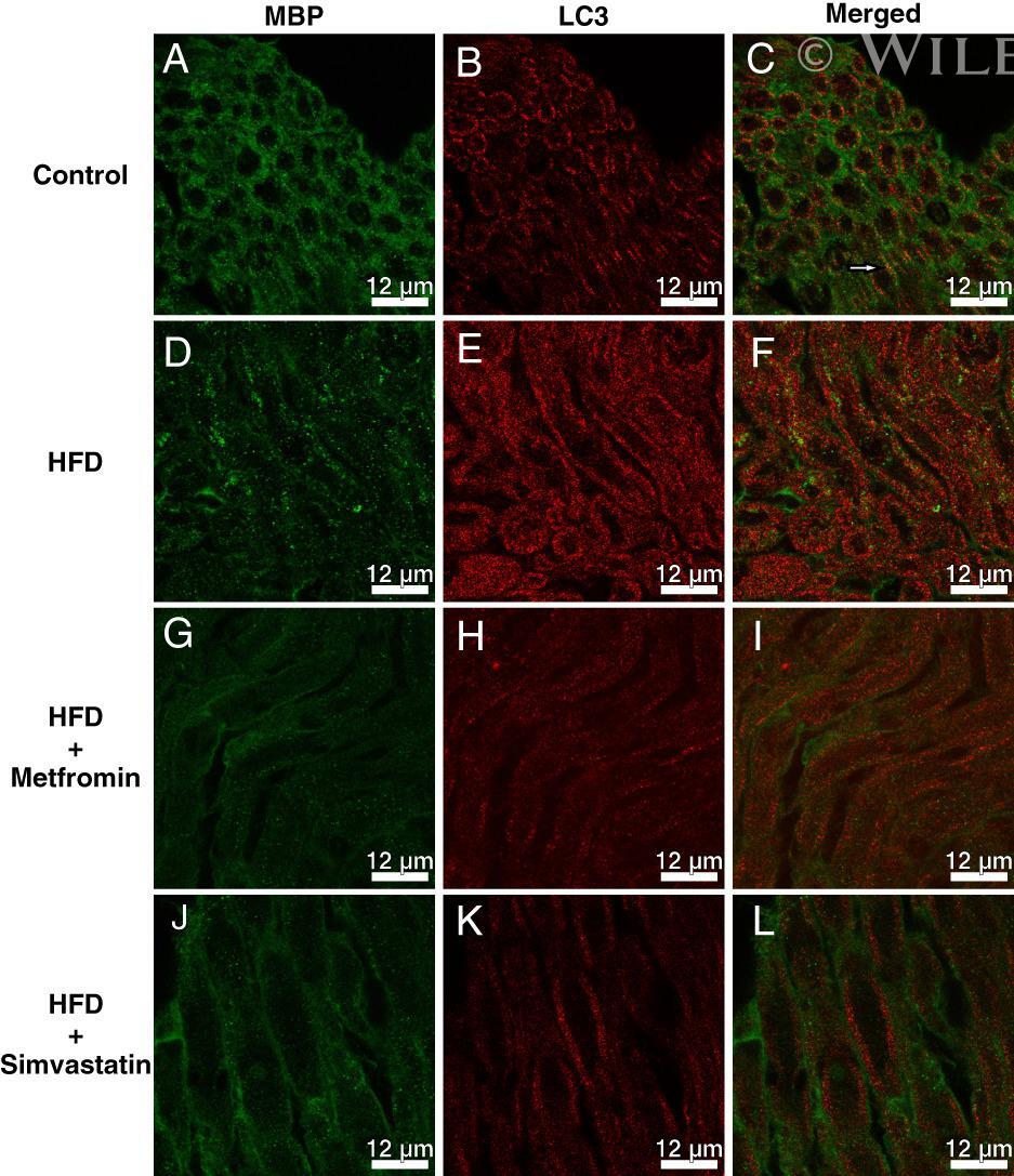
Metformin exacerbates and simvastatin attenuates myelin damage in high fat diet-fed C57BL/6 J mice.
Edvinsson JCA, Maddahi A, Christiansen IM, Reducha PV, Warfvinge K, Sheykhzade M, Edvinsson L, Haanes KA
The journal of headache and pain 2022 Feb 17;23(1):26
The journal of headache and pain 2022 Feb 17;23(1):26
Metformin exacerbates and simvastatin attenuates myelin damage in high fat diet-fed C57BL/6 J mice.
Ciric D, Martinovic T, Petricevic S, Trajkovic V, Bumbasirevic V, Kravic-Stevovic T
Neuropathology : official journal of the Japanese Society of Neuropathology 2018 Oct;38(5):468-474
Neuropathology : official journal of the Japanese Society of Neuropathology 2018 Oct;38(5):468-474
No comments: Submit comment
Supportive validation
- Submitted by
- Invitrogen Antibodies (provider)
- Main image
- Experimental details
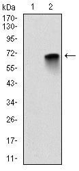
- Western blot analysis of MBP using MBP monoclonal antibody (Product # MA5-15922) in HEK293 (1) and MBP-human IgG Fc transfected HEK293 (2) cell lysate.

- Submitted by
- Invitrogen Antibodies (provider)
- Main image

- Experimental details
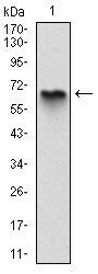
- Western blot analysis of MBP using a MBP monoclonal antibody (Product # MA5-15922) against a human MBP (AA: 1-197) recombinant protein.
- Submitted by
- Invitrogen Antibodies (provider)
- Main image
- Experimental details
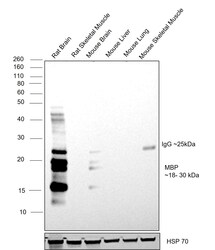
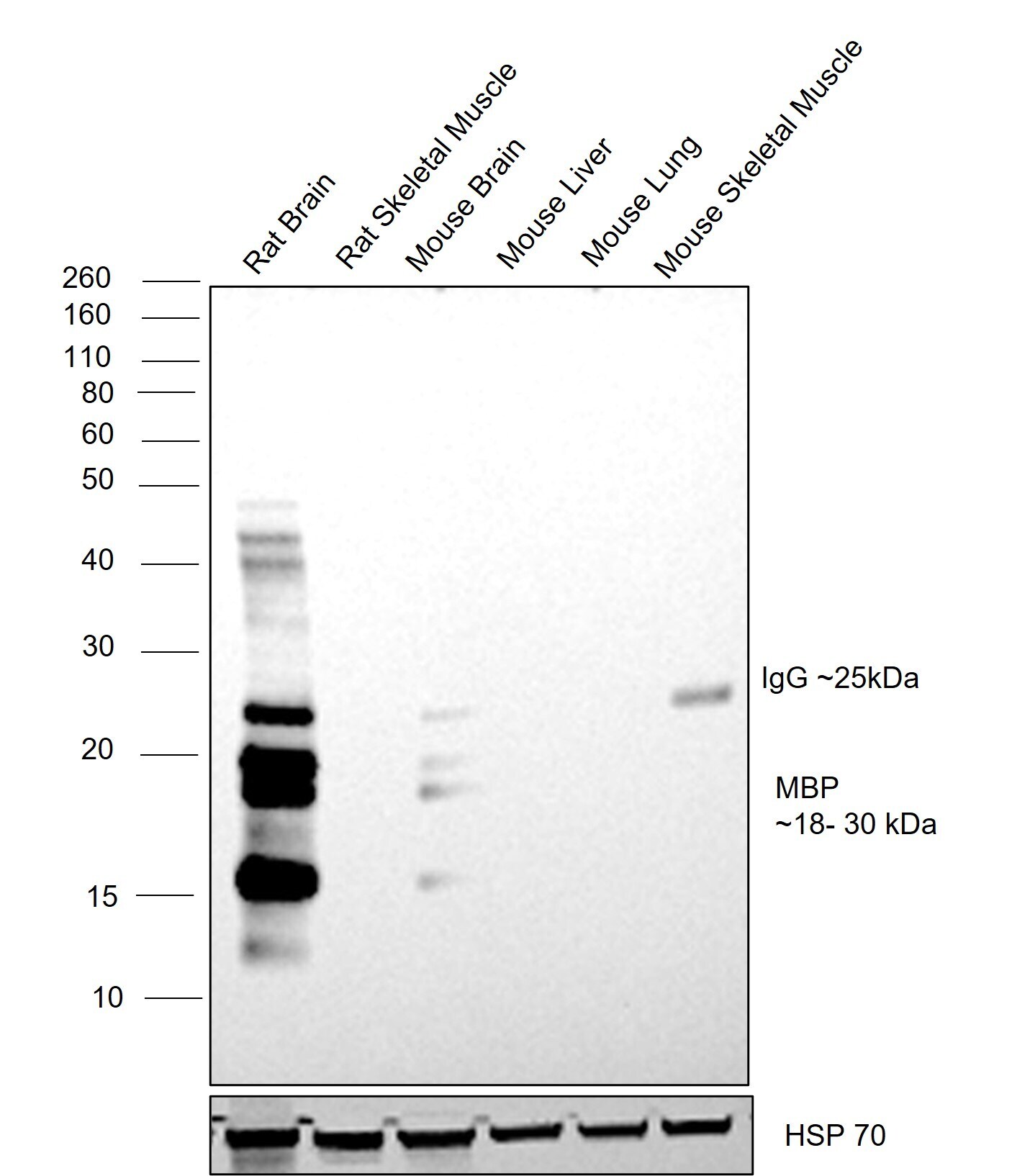
- Western blot was performed using MBP Monoclonal Antibody (2H9) (Product # MA5-15922) and a 18-20 kDa band corresponding to MBP was observed across tissues tested. Immuno-depleted tissue extracts (30 µg lysate) of Rat Brain (Lane 1), Rat Skeletal Muscle (Lane 2), Mouse Brain (Lane 3), Mouse Liver (Lane 4), Mouse Lung (Lane 5), Mouse Skeletal Muscle (Lane 6) were electrophoresed using NuPAGE™ 4-12% Bis-Tris Protein Gel (Product # NP0322BOX). Resolved proteins were then transferred onto a nitrocellulose membrane (Product # IB33001) by iBlot™ 3 Western Blot Transfer Device (Product # IB31001). The blot was probed with the primary antibody (1:2,000) and detected by chemiluminescence with Goat anti-Mouse IgG (H+L) Superclonal™ Recombinant Secondary Antibody, HRP (Product # A28177, 1:20,000) using the iBright™ FL1500 Imaging System (Product # A44115). Chemiluminescent detection was performed using SuperSignal™ West Pico PLUS Chemiluminescent Substrate (Product # 34580). A 25 kDa band corresponding to IgG light chain was observed in mouse tissue lysates.
Supportive validation
- Submitted by
- Invitrogen Antibodies (provider)
- Main image

- Experimental details
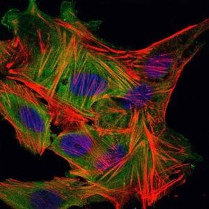
- Immunofluorescence analysis of MSCS cells using MBP monoclonal antibody (Product # MA5-15922) (Green). Blue: DRAQ5 fluorescent DNA dye. Red: actin filaments have been labeled with phalloidin.
- Submitted by
- Invitrogen Antibodies (provider)
- Main image

- Experimental details
- Immunofluorescence analysis of MSCS cells using MBP monoclonal antibody (Product # MA5-15922) (Green). Blue: DRAQ5 fluorescent DNA dye. Red: actin filaments have been labeled with phalloidin.

Supportive validation
- Submitted by
- Invitrogen Antibodies (provider)
- Main image
- Experimental details
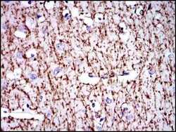
- Immunohistochemical analysis of paraffin-embedded brain tissues using MBP monoclonal antibody (Product # MA5-15922) followed with DAB staining.
- Submitted by
- Invitrogen Antibodies (provider)
- Main image
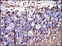
- Experimental details
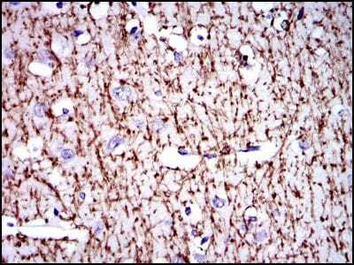
- Immunohistochemical analysis of paraffin-embedded cerebellum tissues using MBP monoclonal antibody (Product # MA5-15922) followed with DAB staining.

Supportive validation
- Submitted by
- Invitrogen Antibodies (provider)
- Main image

- Experimental details
- Flow cytometric analysis of HepG2 cells using MBP monoclonal antibody (Product # MA5-15922) (green) and negative control (red).
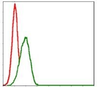
- Submitted by
- Invitrogen Antibodies (provider)
- Main image
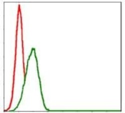
- Experimental details
- Flow cytometric analysis of HepG2 cells using MBP monoclonal antibody (Product # MA5-15922) (green) and negative control (red).

Supportive validation
- Submitted by
- Invitrogen Antibodies (provider)
- Main image

- Experimental details
- Representative confocal lazer microscopic images of double labeled immunofluorescence-stained sciatic nerve sections for MBP (green) and LC3 (red). White arrow indicates yellow colocalization of MBP and LC3.
- Submitted by
- Invitrogen Antibodies (provider)
- Main image
- Experimental details
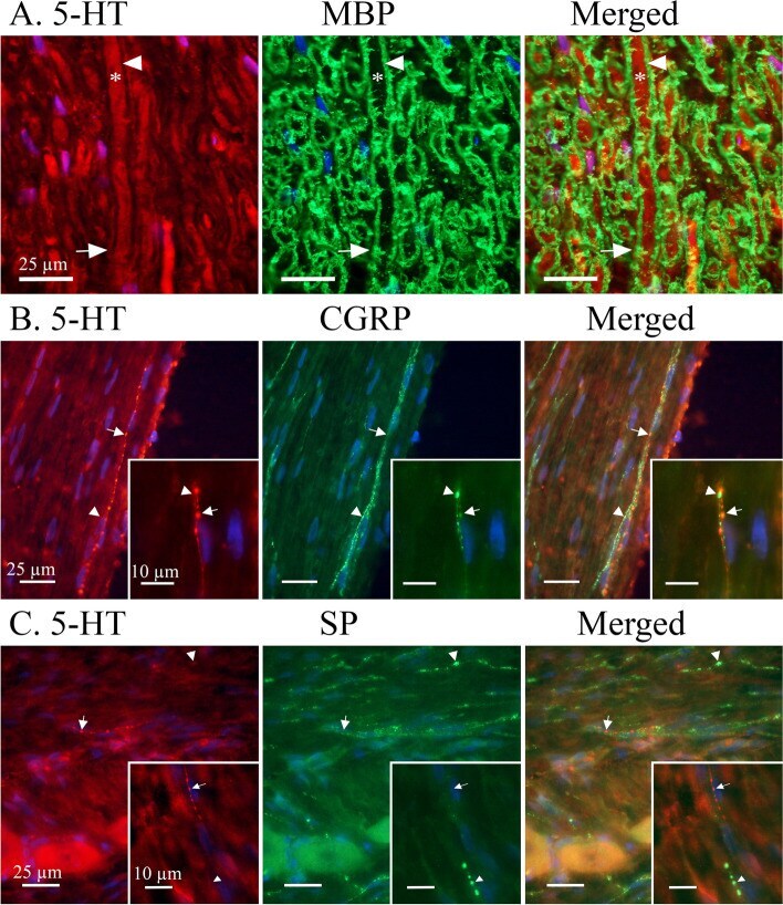
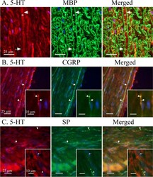
- Expression of 5-HT in relation to myelin basic protein (MBP), SP and CGRP. A MBP showed a distinct staining of myelin sheath flanking cross-sectioned Adelta-fibre axons. A robust MBP-ir was observed (Arrowhead) between the Adelta-fibre axon and Schwann cell neurolemma. An irregular, and sometimes faint, expression of 5-HT could be observed in Adelta-fibre axons (Asterix) and just outside the myelin sheath (Arrow), indicative for the neurolemma of the Schwann cell containing its cytoplasm. B A minority of observed C-fibres expressed 5-HT-ir, these were more frequently found in proximity to Redlich-Obersteiner's zone. CGRP-ir was more abundantly expressed in C-fibres and did not co-localize with 5-HT. Arrows mark 5-HT positive C-fibre boutons and arrowheads mark CGRP positive C-fibre boutons. Insert: Two thin C-fibres entwining and differentially expressing either CGRP or 5-HT. C Similarly, SP could be found concentrated to C-fibre boutons (Arrowhead) throughout the TG. The few 5-HT positive C-fibres (Arrow) were not found to co-localize with SP positive C-fibres. Insert: A thin C-fibre expressing 5-HT-ir (Arrow) above a seemingly larger SP positive C-fibre (Arrowhead)