 Explore
 Validate
 Learn
Learn Western blot
Antibody data
- Antibody Data
- Antigen structure
- References [2]
- Comments [0]
- Validations
- Western blot [2]
- Immunohistochemistry [1]
- Flow cytometry [2]
- Other assay [1]
Submit
Validation data
Reference
Comment
Report error
- Product number
- PA5-13204 - Provider product page

- Provider
- Invitrogen Antibodies
- Product name
- NRG1 Polyclonal Antibody
- Antibody type
- Polyclonal
- Antigen
- Synthetic peptide
- Reactivity
- Human
- Host
- Rabbit
- Isotype
- IgG
- Vial size
- 400 μL
- Concentration
- 0.45 mg/mL
- Storage
- Store at 4°C short term. For long term storage, store at -20°C, avoiding freeze/thaw cycles.
Submitted references Lung injury in axolotl salamanders induces an organ-wide proliferation response.
Targeting HER2 in patient-derived xenograft ovarian cancer models sensitizes tumors to chemotherapy.
Jensen TB, Giunta P, Schultz NG, Griffiths JM, Duerr TJ, Kyeremateng Y, Wong H, Adesina A, Monaghan JR
Developmental dynamics : an official publication of the American Association of Anatomists 2021 Jun;250(6):866-879
Developmental dynamics : an official publication of the American Association of Anatomists 2021 Jun;250(6):866-879
Targeting HER2 in patient-derived xenograft ovarian cancer models sensitizes tumors to chemotherapy.
Harris FR, Zhang P, Yang L, Hou X, Leventakos K, Weroha SJ, Vasmatzis G, Kovtun IV
Molecular oncology 2019 Feb;13(2):132-152
Molecular oncology 2019 Feb;13(2):132-152
No comments: Submit comment
Supportive validation
- Submitted by
- Invitrogen Antibodies (provider)
- Main image

- Experimental details
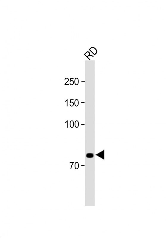
- Western blot analysis of NRG1 in RD whole cell lysate. Samples were incubated with NRG1 polyclonal antibody (Product # PA5-13204) using a dilution of 1:1,000 followed by Goat Anti-Rabbit IgG, (H+L), Peroxidase conjugated at a dilution of 1:15,000. Lysates/proteins: 20 µg per lane. Observed band size: 71 KDa. Blocking/Dilution buffer: 5% NFDM/TBST.
- Submitted by
- Invitrogen Antibodies (provider)
- Main image
- Experimental details
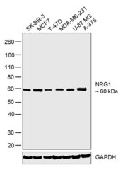
- Western blot was performed using Anti-NRG1 Polyclonal Antibody (Product # PA5-13204) and a 60 kDa band corresponding to NRG1 was observed across the cell lines tested. Whole cell extracts (30 µg) of SK-BR-3 (Lane 1), MCF7 (Lane 2), T47D (Lane 3), MDA-MB-231 (Lane 4), U-87 MG (Lane 5) and A-375 (Lane 6) were electrophoresed using Novex® NuPAGE® 4-12 % Bis-Tris gel (Product # NP0322BOX). Resolved proteins were then transferred onto a nitrocellulose membrane (Product # IB23001) by iBlot® 2 Dry Blotting System (Product # IB21001). The blot was probed with the primary antibody (1:1,000 dilution) and detected by chemiluminescence with Goat anti-Rabbit IgG (Heavy Chain), Superclonal™ Recombinant Secondary Antibody, HRP (Product # A27036, 1:4,000 dilution) using the iBright FL 1000 (Product # A32752). Chemiluminescent detection was performed using Novex® ECL Chemiluminescent Substrate Reagent Kit (Product # WP20005).

Supportive validation
- Submitted by
- Invitrogen Antibodies (provider)
- Main image

- Experimental details
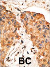
- Immunohistochemistry analysis of NRG1 in formalin-fixed and paraffin-embedded human cancer tissue. Samples were incubated with NRG1 polyclonal antibody (Product # PA5-13204) which was peroxidase-conjugated to the secondary antibody, followed by DAB staining. This data demonstrates the use of this antibody for immunohistochemistry; clinical relevance has not been evaluated. BC = breast carcinoma; HC = hepatocarcinoma.
Supportive validation
- Submitted by
- Invitrogen Antibodies (provider)
- Main image
- Experimental details
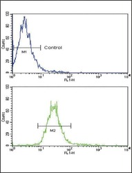
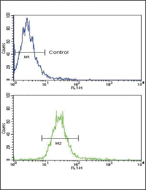
- Flow cytometry analysis of NCI-H460 cells using a NRG1 polyclonal antibody (Product # PA5-13204) (bottom), compared to a negative control cell (top) at a dilution of 1:10-50, followed by a FITC-conjugated goat anti-rabbit antibody
- Submitted by
- Invitrogen Antibodies (provider)
- Main image
- Experimental details
- Flow cytometry of NRG1 in NCI-H460 cells (bottom histogram). Samples were incubated with NRG1 polyclonal antibody (Product # PA5-13204) followed by FITC-conjugated goat-anti-rabbit secondary antibody. Negative control (top histogram).

Supportive validation
- Submitted by
- Invitrogen Antibodies (provider)
- Main image
- Experimental details
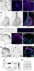
- 4 FIGURE Expression of genes associated with Nrg1/ErbB signaling. (A) HCR FISH for nrg1 in the IX/X cranial nerve and in the spinal cord. Scale bar = 500 um. Inset = 100 um. (B) Immunohistochemistry of Nrg1 and acetylated beta-Tubulin staining cell bodies and axons of peripheral nerves. Scale bar = 100 um. (C-C') Immunohistochemistry of Nrg1 in the injured lung 21 dpa. Notice expression in ciliated cells and smooth muscle cells. (C'') No primary control immunohistochemical stain. Scale bar in C = 250 um. Scale bar in C' = 50 um. Scale bar in C'' = 100 um. (D) HCR FISH of nrg1 and EdU staining in the injured lung 21 dpa. Notice proliferation in both epithelial and mesenchymal cell types. (E) Heat map of qPCR fold increases at both 7 dpa (n = 3) and 21 dpa (n = 4) for the left (injured) and right (contralateral) lungs. Red asterisks denote significant upregulation of mRNA products ( P < 0.05), while orange asterisks denote trending toward significance ( P < 0.10). (E') Table of significant fold changes ( P < 0.05) for the qPCR results with fold changes indicated. IX/X, IX/X cranial nerve ganglion; o, optic cup; sc, spinal cord
