Explore
 Validate
 Learn
Learn Western blot
 Immunocytochemistry
Antibody data
- Antibody Data
- Antigen structure
- References [4]
- Comments [0]
- Validations
- Immunocytochemistry [1]
- Immunohistochemistry [1]
Submit
Validation data
Reference
Comment
Report error
- Product number
- HPA028275 - Provider product page

- Provider
- Atlas Antibodies
- Proper citation
- Atlas Antibodies Cat#HPA028275, RRID:AB_10600909
- Product name
- Anti-FABP1
- Antibody type
- Polyclonal
- Description
- Polyclonal Antibody against Human FABP1, Gene description: fatty acid binding protein 1, liver, Alternative Gene Names: L-FABP, Validated applications: WB, IHC, ICC, Uniprot ID: P07148, Storage: Store at +4°C for short term storage. Long time storage is recommended at -20°C.
- Reactivity
- Human, Mouse, Rat
- Host
- Rabbit
- Conjugate
- Unconjugated
- Isotype
- IgG
- Vial size
- 100 µl
- Concentration
- 0.1 mg/ml
- Storage
- Store at +4°C for short term storage. Long time storage is recommended at -20°C.
- Handling
- The antibody solution should be gently mixed before use.
Submitted references The role of altered lipid composition and distribution in liver fibrosis revealed by multimodal nonlinear optical microscopy
ACSS2 promotes systemic fat storage and utilization through selective regulation of genes involved in lipid metabolism
Elevated levels of circulating CDH5 and FABP1 in association with human drug-induced liver injury.
Hepatitis B Virus X Protein Induces Hepatic Steatosis by Enhancing the Expression of Liver Fatty Acid Binding Protein
Jia H, Liu J, Fang T, Zhou Z, Li R, Yin W, Qian Y, Wang Q, Zhou W, Liu C, Sun D, Chen X, Ouyang Z, Dong J, Wang Y, Yue S
Science Advances 2023;9(2)
Science Advances 2023;9(2)
ACSS2 promotes systemic fat storage and utilization through selective regulation of genes involved in lipid metabolism
Huang Z, Zhang M, Plec A, Estill S, Cai L, Repa J, McKnight S, Tu B
Proceedings of the National Academy of Sciences 2018;115(40)
Proceedings of the National Academy of Sciences 2018;115(40)
Elevated levels of circulating CDH5 and FABP1 in association with human drug-induced liver injury.
Mikus M, Drobin K, Gry M, Bachmann J, Lindberg J, Yimer G, Aklillu E, Makonnen E, Aderaye G, Roach J, Fier I, Kampf C, Göpfert J, Perazzo H, Poynard T, Stephens C, Andrade RJ, Lucena MI, Arber N, Uhlén M, Watkins PB, Schwenk JM, Nilsson P, Schuppe-Koistinen I
Liver international : official journal of the International Association for the Study of the Liver 2017 Jan;37(1):132-140
Liver international : official journal of the International Association for the Study of the Liver 2017 Jan;37(1):132-140
Hepatitis B Virus X Protein Induces Hepatic Steatosis by Enhancing the Expression of Liver Fatty Acid Binding Protein
Wu Y, Peng X, Zhu Y, Yan X, Chen W, Lin X, Sandri-Goldin R
Journal of Virology 2016;90(4):1729-1740
Journal of Virology 2016;90(4):1729-1740
No comments: Submit comment
Supportive validation
- Submitted by
- Atlas Antibodies (provider)
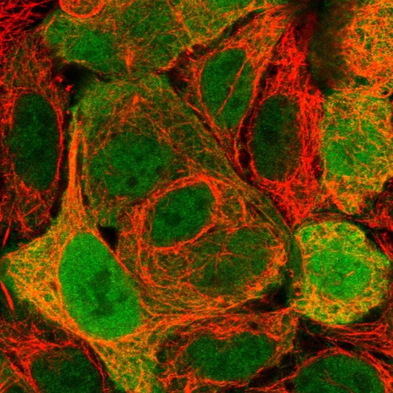
- Main image
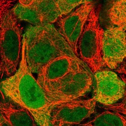
- Experimental details
- Immunofluorescent staining of human cell line CACO-2 shows localization to nucleoplasm & cytosol.
- Sample type
- Human
Supportive validation
- Submitted by
- Atlas Antibodies (provider)
- Enhanced method
- Orthogonal validation
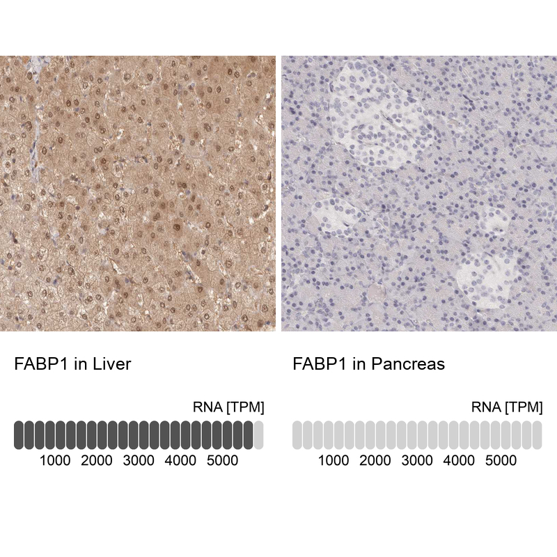
- Main image
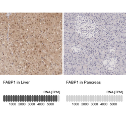
- Experimental details
- Immunohistochemistry analysis in human liver and pancreas tissues using HPA028275 antibody. Corresponding FABP1 RNA-seq data are presented for the same tissues.
- Sample type
- Human
- Protocol
- Protocol