 Explore
 Validate
 Learn
Learn Western blot
 Immunohistochemistry
Antibody data
- Antibody Data
- Antigen structure
- References [1]
- Comments [0]
- Validations
- Western blot [3]
Submit
Validation data
Reference
Comment
Report error
- Product number
- MAB1565 - Provider product page

- Provider
- R&D Systems
- Product name
- Mouse/Rat FABP1/L-FABP Antibody
- Antibody type
- Monoclonal
- Description
- Protein A or G purified from hybridoma culture supernatant. Detects rat FABP1/L-FABP in direct ELISAs and Western blots. In direct ELISAs and Western blots no cross-reactivity with recombinant rat FABP2, recombinant human FABP3, recombinant mouse (rm) FABP4, or rmFABP5 is observed.
- Reactivity
- Mouse, Rat
- Host
- Mouse
- Conjugate
- Unconjugated
- Antigen sequence
P02692- Isotype
- IgG
- Antibody clone number
- 220119
- Vial size
- 100 ug
- Concentration
- LYOPH
- Storage
- Use a manual defrost freezer and avoid repeated freeze-thaw cycles. 12 months from date of receipt, -20 to -70 °C as supplied. 1 month, 2 to 8 °C under sterile conditions after reconstitution. 6 months, -20 to -70 °C under sterile conditions after reconstitution.
Submitted references Helper-dependent adenovirus-mediated short hairpin RNA expression in the liver activates the interferon response.
Witting SR, Brown M, Saxena R, Nabinger S, Morral N
The Journal of biological chemistry 2008 Jan 25;283(4):2120-8
The Journal of biological chemistry 2008 Jan 25;283(4):2120-8
No comments: Submit comment
Supportive validation
- Submitted by
- R&D Systems (provider)
- Main image
- Experimental details
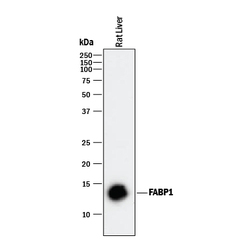
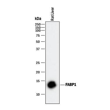
- Detection of Rat FABP1/L-FABP by Western Blot. Western blot shows lysate of rat liver tissue. PVDF membrane was probed with 2 µg/mL of Mouse Anti-Mouse/Rat FABP1/L-FABP Monoclonal Antibody (Catalog # MAB1565) followed by HRP-conjugated Anti-Mouse IgG Secondary Antibody (Catalog # HAF018). A specific band was detected for FABP1/L-FABP at approximately 13 kDa (as indicated). This experiment was conducted under reducing conditions and using Immunoblot Buffer Group 1.
- Submitted by
- R&D Systems (provider)
- Main image
- Experimental details
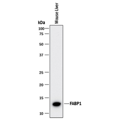
- Detection of Mouse FABP1/L-FABP by Western Blot. Western blot shows lysates of mouse liver tissue. PVDF membrane was probed with 2 µg/mL of Mouse Anti-Mouse/ Rat FABP1/L-FABP Monoclonal Antibody (Catalog # MAB1565) followed by HRP-conjugated Anti-Mouse IgG Secondary Antibody (Catalog # HAF018). A specific band was detected for FABP1/L-FABP at approximately 13 kDa (as indicated). This experiment was conducted under reducing conditions and using Immunoblot Buffer Group 1.

- Submitted by
- R&D Systems (provider)
- Main image
- Experimental details
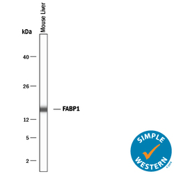
- Detection of Mouse FABP1/L-FABP by Simple WesternTM. Simple Western lane view shows lysates of mouse liver tissue, loaded at 0.2 mg/mL. A specific band was detected for FABP1/L-FABP at approximately 16 kDa (as indicated) using 10 µg/mL of Mouse Anti-Mouse/Rat FABP1/L-FABP Monoclonal Antibody (Catalog # MAB1565). This experiment was conducted under reducing conditions and using the 12-230 kDa separation system.
