Explore
 Validate
 Learn
Learn Western blot
Antibody data
- Antibody Data
- Antigen structure
- References [0]
- Comments [0]
- Validations
- Western blot [4]
- Immunohistochemistry [1]
Submit
Validation data
Reference
Comment
Report error
- Product number
- MA5-24055 - Provider product page

- Provider
- Invitrogen Antibodies
- Product name
- FABP1 Monoclonal Antibody (328607)
- Antibody type
- Monoclonal
- Antigen
- Recombinant full-length protein
- Description
- In direct ELISAs, no cross-reactivity with recombinant human FABP2, -3, -4, -5, -6, -7, or recombinant mouse FABP9 is observed.
- Antibody clone number
- 328607
- Concentration
- 0.5 mg/mL
No comments: Submit comment
Supportive validation
- Submitted by
- Invitrogen Antibodies (provider)
- Main image

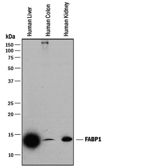
- Experimental details
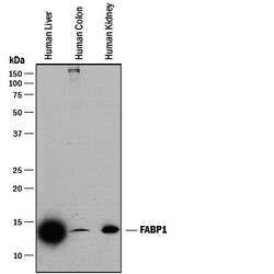
- Western blot analysis from lysates of human liver tissue, human colon tissue, and human kidney tissue. PVDF membrane was probed with 0.25 µg/mL of mouse Anti-human FABP1/L-FABP Monoclonal Antibody (Product # MA5-24055) followed by HRP-conjugated Anti-mouse IgG Secondary Antibody. A specific band was detected for FABP1/L-FABP at approximately 14 kDa (as indicated). This experiment was conducted under reducing conditions.
- Submitted by
- Invitrogen Antibodies (provider)
- Main image
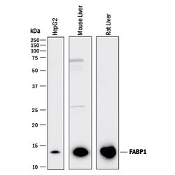
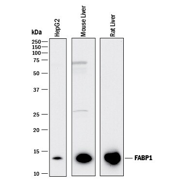
- Experimental details
- Western blot analysis of FABP1 in HepG2 human hepatocellular carcinoma cell line, mouse liver tissue, and rat liver tissue. Samples were incubated in FABP1 monoclonal antibody (Product # MA5-24055) using a dilution of 0.25 µg/mL followed by a HRP-conjugated Anti-Mouse IgG secondary antibody. A specific band was detected for FABP1/L‚FABP at approximately 14 kDa (as indicated). This experiment was conducted under reducing conditions.
- Submitted by
- Invitrogen Antibodies (provider)
- Main image
- Experimental details
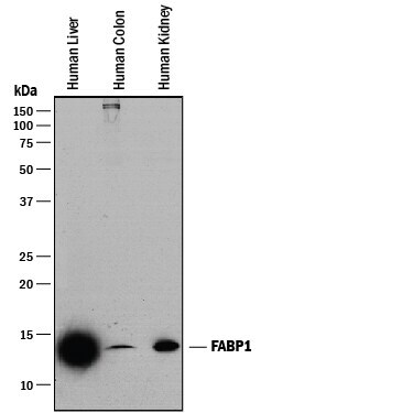
- Western blot analysis of FABP1 in human liver tissue, human colon tissue, and human kidney tissue. Samples were incubated in FABP1 monoclonal antibody (Product # MA5-24055) using a dilution of 0.25 µg/mL followed by a HRP-conjugated Anti-Mouse IgG secondary antibody. A specific band was detected for FABP1/L‚FABP at approximately 14 kDa (as indicated). This experiment was conducted under reducing conditions.
- Submitted by
- Invitrogen Antibodies (provider)
- Main image
- Experimental details
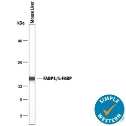
- Western blot analysis of FABP1 in 0.2 mg/mL lysates of mouse liver tissue. Samples were incubated in FABP1 monoclonal antibody (Product # MA5-24055) using a dilution of 5 µg/mL. A specific band was detected for FABP1/L‚FABP at approximately 16 kDa (as indicated). This experiment was conducted under reducing conditions and using the 12-230 kDa separation system.

Supportive validation
- Submitted by
- Invitrogen Antibodies (provider)
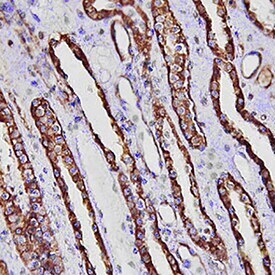
- Main image

- Experimental details
- Immunohistochemical analysis of FABP1 in immersion fixed paraffin-embedded sections of human kidney. Samples were incubated in FABP1 monoclonal antibody (Product # MA5-24055) using a dilution of 15 µg/mL overnight at 4 °C. Tissue was stained using the Anti-Mouse HRP-DAB Cell & Tissue Staining Kit (brown) and counterstained with hematoxylin (blue). Specific staining was localized to convoluted tubules.