Explore
 Validate
 Learn
Learn Western blot
Antibody data
- Antibody Data
- Antigen structure
- References [4]
- Comments [0]
- Validations
- Western blot [1]
- Immunohistochemistry [1]
Submit
Validation data
Reference
Comment
Report error
- Product number
- SC-15378 - Provider product page

- Provider
- Santa Cruz Biotechnology
- Proper citation
- Santa Cruz Biotechnology Cat#sc-15378, RRID:AB_2100047
- Product name
- Anti-EMD
- Antibody type
- Polyclonal
- Antigen
- Recombinant full-length protein
- Reactivity
- Human
- Host
- Rabbit
Submitted references Barrier-to-autointegration factor proteome reveals chromatin-regulatory partners.
Tyrosine phosphorylation of nuclear-membrane protein emerin by Src, Abl and other kinases.
Emerin is hyperphosphorylated and redistributed in herpes simplex virus type 1-infected cells in a manner dependent on both UL34 and US3.
Homozygous and compound heterozygous mutations in ZMPSTE24 cause the laminopathy restrictive dermopathy.
Montes de Oca R, Shoemaker CJ, Gucek M, Cole RN, Wilson KL
PloS one 2009 Sep 16;4(9):e7050
PloS one 2009 Sep 16;4(9):e7050
Tyrosine phosphorylation of nuclear-membrane protein emerin by Src, Abl and other kinases.
Tifft KE, Bradbury KA, Wilson KL
Journal of cell science 2009 Oct 15;122(Pt 20):3780-90
Journal of cell science 2009 Oct 15;122(Pt 20):3780-90
Emerin is hyperphosphorylated and redistributed in herpes simplex virus type 1-infected cells in a manner dependent on both UL34 and US3.
Leach N, Bjerke SL, Christensen DK, Bouchard JM, Mou F, Park R, Baines J, Haraguchi T, Roller RJ
Journal of virology 2007 Oct;81(19):10792-803
Journal of virology 2007 Oct;81(19):10792-803
Homozygous and compound heterozygous mutations in ZMPSTE24 cause the laminopathy restrictive dermopathy.
Moulson CL, Go G, Gardner JM, van der Wal AC, Smitt JH, van Hagen JM, Miner JH
The Journal of investigative dermatology 2005 Nov;125(5):913-9
The Journal of investigative dermatology 2005 Nov;125(5):913-9
No comments: Submit comment
Supportive validation
- Submitted by
- per
- Main image

- Experimental details
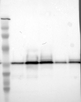
- Western blot analysis of antibody specificity using a routine panel composed of IgG/HSA-depleted human plasma and protein lysates from selected human tissues and cell lines.
- Validation comment
- Single band corresponding to the predicted size in kDa (+/-20%).
- Primary Ab dilution
- 1:500
- Secondary Ab dilution
- 1:3000
- Lane 1
- Marker [kDa]: 206, 113, 82, 49, 32, 26, 17.8
- Lane 2
- RT-4
- Lane 3
- U-251MG sp
- Lane 4
- A-431
- Lane 5
- Liver
- Lane 6
- Tonsil
- Theoretical target weight
- [kDa] 25
Supportive validation
- Submitted by
- per
- Main image
- Experimental details
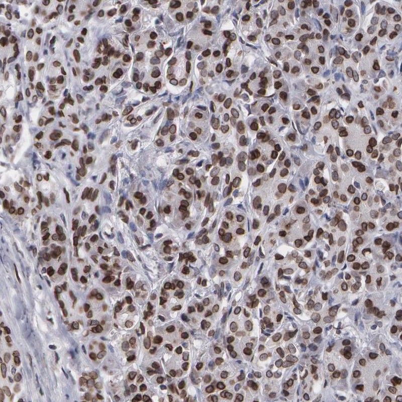
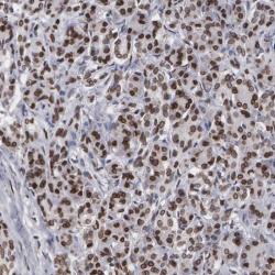
- Immunohistochemical staining of human pancreas shows strong nuclear positivity in exocrine glandular cells.
- Validation comment
- Two independent antibodies targeting one protein yielding similar staining patterns. Staining pattern consistent with experimental and/or bioinformatic data.