Antibody data
- Antibody Data
- Antigen structure
- References [0]
- Comments [0]
- Validations
- Western blot [3]
- Immunocytochemistry [5]
- Immunohistochemistry [5]
Submit
Validation data
Reference
Comment
Report error
- Product number
- AMAb90562 - Provider product page
- Provider
- Atlas Antibodies
- Proper citation
- Atlas Antibodies Cat#AMAb90562, RRID:AB_2665587
- Product name
- Anti-EMD
- Antibody type
- Monoclonal
- Reactivity
- Human
- Host
- Mouse
- Conjugate
- Unconjugated
- Antigen sequence
ASSYSFSDLNSTRGDADMYDLPKKEDALLYQSKGY
NDDYYEESYFTTRTYGEPESAGPSRAVRQSVTSFP
DADAFHHQVHDDDLLSSSEEECKDRERPMYGRDSA
CQSITHY- Epitope
- Binds to an epitope located within the peptide sequence QSKGYNDDYY as determined by overlapping synthetic peptides.
- Isotype
- IgG
- Antibody clone number
- CL0203
- Vial size
- 100 µl
- Storage
- Store at +4°C for short term storage. Long time storage is recommended at -20°C.
No comments: Submit comment
Enhanced validation
- Submitted by
- Atlas Antibodies (provider)
- Enhanced method
- Genetic validation
- Main image
- Experimental details
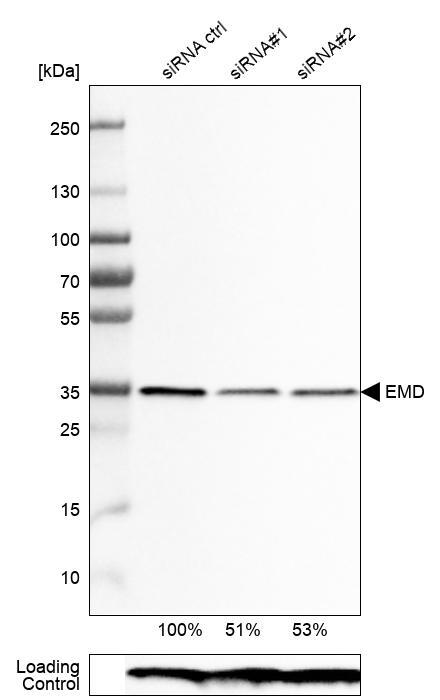
- Western blot analysis in RT-4 cells transfected with control siRNA, target specific siRNA probe #1 and #2, using Anti-EMD antibody. Remaining relative intensity is presented. Loading control: Anti-PPIB.
- Submitted by
- Atlas Antibodies (provider)
- Main image
- Experimental details
- Lane 1: Marker [kDa]Lane 2: RT-4
- Submitted by
- Atlas Antibodies (provider)
- Main image
- Experimental details
- Western blot analysis of extracts from RT-4 cells, transfected with: control siRNA, target specific siRNA probe #1, target specific siRNA probe #2, using Anti-EMD monoclonal antibody. Downregulation of antibody signal confirms target specificity. Remaining % intensity, relative control lane, is indicated. Anti-PPIB monoclonal antibody was used as loading control.
Supportive validation
- Submitted by
- Atlas Antibodies (provider)
- Main image
- Experimental details
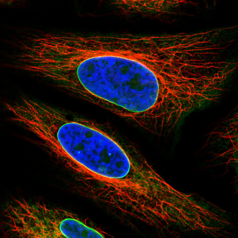
- Immunofluorescence staining in HeLa cell line with Anti-EMD monoclonal antibody, shows a distinct nuclear membranous pattern with a weaker additional ER staining in green. Microtubule- and nuclear probes are visualized in red and blue respectively (where available).
- Sample type
- HUMAN
- Submitted by
- Atlas Antibodies (provider)
- Main image
- Experimental details
- Immunofluorescence staining in A431 cell line with Anti-EMD monoclonal antibody, shows a distinct nuclear membranous pattern with a weaker additional ER staining in green. Microtubule- and nuclear probes are visualized in red and blue respectively (where available).
- Sample type
- HUMAN
- Submitted by
- Atlas Antibodies (provider)
- Main image
- Experimental details
- Immunofluorescence staining in MCF7 cell line with Anti-EMD monoclonal antibody, shows a distinct nuclear membranous pattern with a weaker additional ER staining in green. Microtubule- and nuclear probes are visualized in red and blue respectively (where available).
- Sample type
- HUMAN
- Submitted by
- Atlas Antibodies (provider)
- Main image
- Experimental details
- Immunofluorescence staining in U2OS cell line with Anti-EMD monoclonal antibody, shows a distinct nuclear membranous pattern with a weaker additional ER staining in green. Microtubule- and nuclear probes are visualized in red and blue respectively (where available).
- Sample type
- HUMAN
- Submitted by
- Atlas Antibodies (provider)
- Main image
- Experimental details
- Immunofluorescence staining in U251 cell line with Anti-EMD monoclonal antibody, shows a distinct nuclear membranous pattern with a weaker additional ER staining in green. Microtubule- and nuclear probes are visualized in red and blue respectively (where available).
- Sample type
- HUMAN
Supportive validation
- Submitted by
- Atlas Antibodies (provider)
- Main image
- Experimental details
- Immunohistochemical staining of human skin shows strong nuclear membrane positivity in the epidermis cells.
- Submitted by
- Atlas Antibodies (provider)
- Main image
- Experimental details
- Immunohistochemical staining of human prostate shows strong nuclear membrane immunoreactivityy in glandular cells.
- Submitted by
- Atlas Antibodies (provider)
- Main image
- Experimental details
- Immunohistochemical staining of human placenta shows strong nuclear membrane positivity in the trophoblast.
- Submitted by
- Atlas Antibodies (provider)
- Main image
- Experimental details
- Immunohistochemical staining of human testis shows nuclear membrane immunoreactivity.
- Submitted by
- Atlas Antibodies (provider)
- Main image
- Experimental details
- Immunohistochemical staining of human kidney shows nuclear membrane positivity in renal tubules.