Explore
 Validate
 Learn
Learn Western blot
Antibody data
- Antibody Data
- Antigen structure
- References [1]
- Comments [0]
- Validations
- Western blot [5]
- Immunocytochemistry [2]
- Immunohistochemistry [1]
Submit
Validation data
Reference
Comment
Report error
- Product number
- PA5-51424 - Provider product page

- Provider
- Invitrogen Antibodies
- Product name
- Emerin Polyclonal Antibody
- Antibody type
- Polyclonal
- Antigen
- Recombinant full-length protein
- Description
- Immunogen sequence: ASSYSFSDLN STRGDADMYD LPKKEDALLY QSKGYNDDYY EESYFTTRTY GEPESAGPSR AVRQSVTSFP DADAFHHQVH DDDLLSSSEE ECKDRERPMY GRDSACQSIT HY
- Concentration
- 0.2 mg/mL
Submitted references The cell-wide web coordinates cellular processes by directing site-specific Ca(2+) flux across cytoplasmic nanocourses.
Duan J, Navarro-Dorado J, Clark JH, Kinnear NP, Meinke P, Schirmer EC, Evans AM
Nature communications 2019 May 24;10(1):2299
Nature communications 2019 May 24;10(1):2299
No comments: Submit comment
Supportive validation
- Submitted by
- Invitrogen Antibodies (provider)
- Main image
- Experimental details
- Western blot analysis of Emerin in RT-4 cells transfected with control siRNA, target specific siRNA probe #1 and #2, using a Emerin Polyclonal Antibody (Product # PA5-51424). Remaining relative intensity is presented. Loading control: Anti-GAPDH.

- Submitted by
- Invitrogen Antibodies (provider)
- Main image
- Experimental details
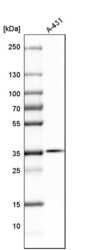
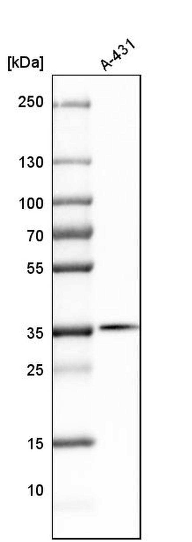
- Western blot analysis of Emerin in human cell line A-431 using a Emerin Polyclonal Antibody (Product # PA5-51424).
- Submitted by
- Invitrogen Antibodies (provider)
- Main image

- Experimental details
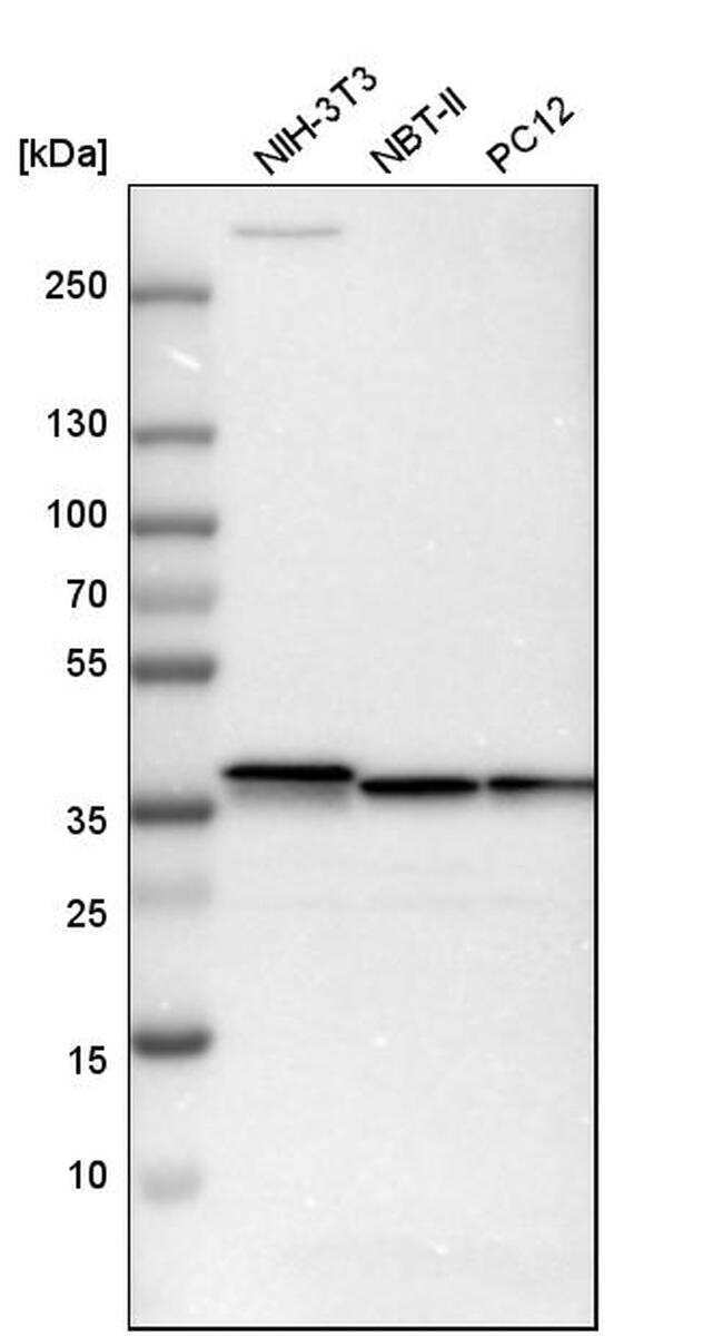
- Western blot analysis of Emerin in mouse cell line NIH-3T3, rat cell line NBT-II and rat cell line pC12 using a Emerin Polyclonal Antibody (Product # PA5-51424).
- Submitted by
- Invitrogen Antibodies (provider)
- Main image
- Experimental details
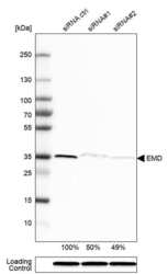
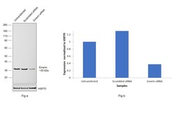
- Knockdown of Emerin was achieved by transfecting HEK-293 with Emerin specific siRNAs (Silencer® select Product # s225840, s4645). Western blot analysis (Fig. a) was performed using Nuclear enriched extracts from the Emerin knockdown cells (lane 3), non-targeting scrambled siRNA transfected cells (lane 2) and untransfected cells (lane 1). The blot was probed with Emerin Polyclonal Antibody (Product # PA5-51424, 0.2 µg/mL) and Goat anti-Rabbit IgG (H+L) Superclonal™ Recombinant Secondary Antibody, HRP (Product # A27036, 1:4000). Densitometric analysis of this western blot is shown in histogram (Fig. b). Decrease in signal upon siRNA mediated knock down confirms that antibody is specific to Emerin.

- Submitted by
- Invitrogen Antibodies (provider)
- Main image
- Experimental details
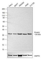
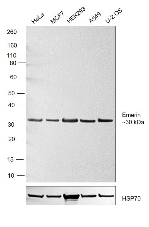
- Western blot was performed using Anti-Emerin Polyclonal Antibody (Product # PA5-51424) and a 29 kDa band corresponding to Emerin was observed across 5 cell lines. Nuclear enriched extracts (30 µg lysate) of HeLa (Lane 1), MCF7 (Lane 2), HEK-293 (Lane 3), A549 (Lane 4), U-2 OS (Lane 5) were electrophoresed using NuPAGE™ 4-12% Bis-Tris Protein Gel (Product # NP0321BOX). Resolved proteins were then transferred onto a Nitrocellulose membrane (Product # IB23002) by iBlot® 2 Dry Blotting System (Product # IB21001). The blot was probed with the primary antibody (0.4 µg/mL) and detected by chemiluminescence with Goat anti-Rabbit IgG (H+L) Superclonal™ Recombinant Secondary Antibody, HRP (Product # A27036, 4000) using the iBright FL 1000 (Product # A32752). Chemiluminescent detection was performed using Novex® ECL Reagent Kit (Product # WP20005).
Supportive validation
- Submitted by
- Invitrogen Antibodies (provider)
- Main image

- Experimental details
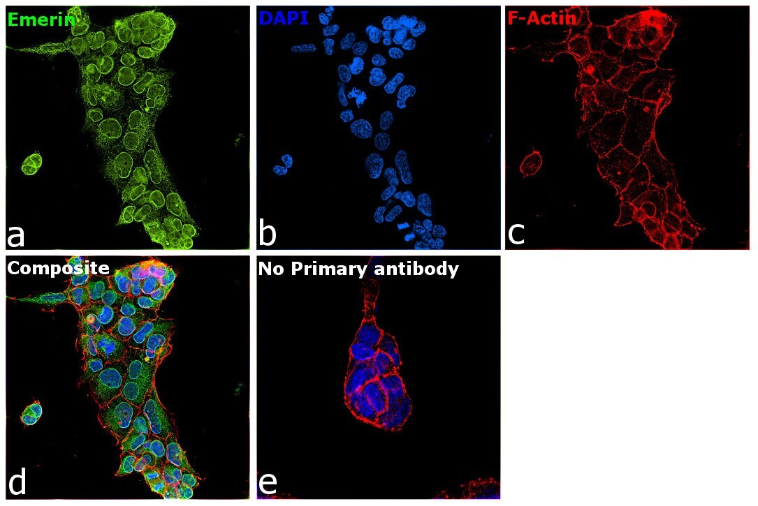
- Immunofluorescent staining of Emerin in human cell line U-251 MG shows positivity in nuclear membrane & endoplasmic reticulum. Samples were probed using an Emerin Polyclonal Antibody (Product # PA5-51424).
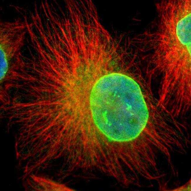
- Submitted by
- Invitrogen Antibodies (provider)
- Main image

- Experimental details
- Immunofluorescence analysis of Emerin was performed using 70% confluent log phase HEK-293 cells. The cells were fixed with 4% paraformaldehyde for 10 minutes, permeabilized with 0.1% Triton™ X-100 for 15 minutes, and blocked with 2% BSA for 45 minutes at room temperature. The cells were labeled with Emerin Polyclonal Antibody (Product # PA5-51424) at 0.2 µg/mL in 0.1% BSA, incubated at 4 degree celsius overnight and then labeled with Goat anti-Rabbit IgG (H+L) Superclonal™ Recombinant Secondary Antibody, Alexa Fluor® 488 conjugate (Product # A27034), (1:2000), for 45 minutes at room temperature (Panel a: Green). Nuclei (Panel b:Blue) were stained with ProLong™ Diamond Antifade Mountant with DAPI (Product # P36962). F-actin (Panel c: Red) was stained with Rhodamine Phalloidin (Product # R415, 1:300). Panel d represents the merged image showing predominantly nuclear membrane localization. Panel e represents control cells with no primary antibody to assess background. The images were captured at 60x magnification.
Supportive validation
- Submitted by
- Invitrogen Antibodies (provider)
- Main image

- Experimental details
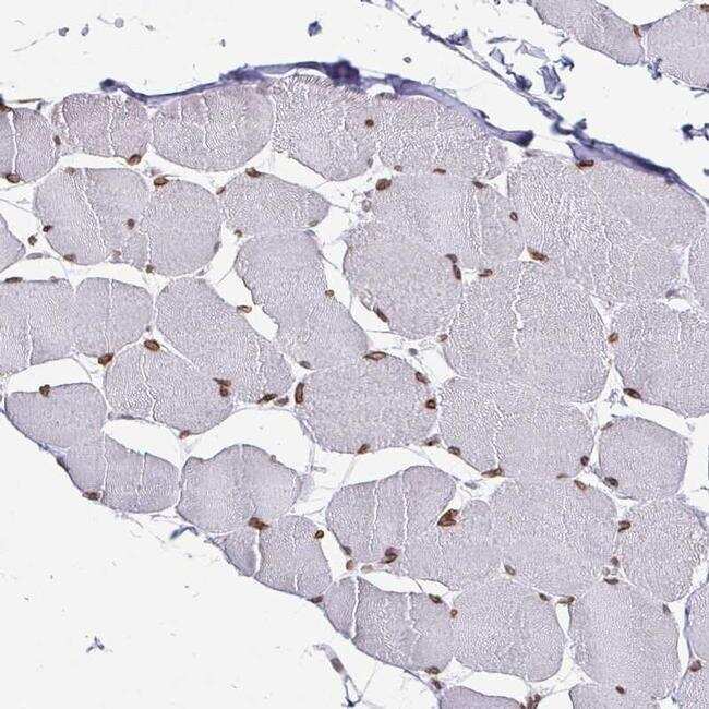
- Immunohistochemical staining of Emerin in human skeletal muscle shows moderate nuclear membrane positivity in myocytes. Samples were probed using an Emerin Polyclonal Antibody (Product # PA5-51424).