Explore
 Validate
 Learn
Learn Western blot
 Immunocytochemistry
Antibody data
- Antibody Data
- Antigen structure
- References [1]
- Comments [0]
- Validations
- Western blot [1]
Submit
Validation data
Reference
Comment
Report error
- Product number
- M00714 - Provider product page

- Provider
- Boster Biological Technology
- Product name
- Anti-Emerin EMD Antibody Picoband™ (monoclonal, 5A10)
- Antibody type
- Monoclonal
- Description
- Mouse IgG monoclonal antibody for Emerin detection. Tested with WB, IHC-P, IHC-F, ICC, FCM in Human.
- Reactivity
- Human
- Host
- Mouse
- Isotype
- IgG
- Antibody clone number
- 5A10
- Vial size
- 100μg/vial
- Concentration
- 0.5-1mg/ml, actual concentration vary by lot. Use suggested dilution ratio to decide dilution procedure.
- Storage
- At -20°C for one year. After reconstitution, at 4°C for one month. It can also be aliquoted and stored frozen at -20°C for a longer time. Avoid repeated freezing and thawing.
- Handling
- Add 0.2ml of distilled water will yield a concentration of 500ug/ml.
Submitted references Collective analysis of the expression and prognosis for LEM-domain proteins in prostate cancer.
He T, Zhang Y, Li X, Liu C, Zhu G, Yin X, Zhang Z, Zhao K, Wang Z, Zhao P, Wang K
World journal of surgical oncology 2022 Jun 2;20(1):174
World journal of surgical oncology 2022 Jun 2;20(1):174
No comments: Submit comment
Supportive validation
- Submitted by
- Boster Biological Technology (provider)
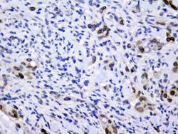
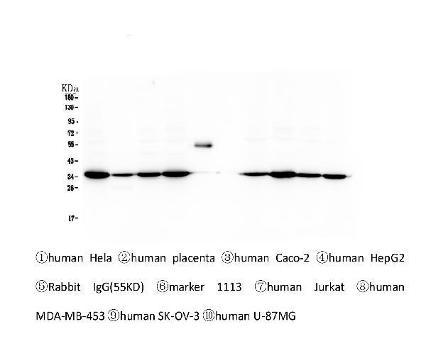
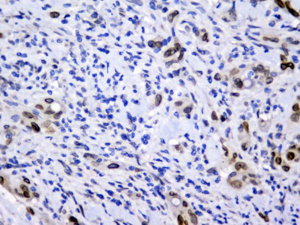
- Main image
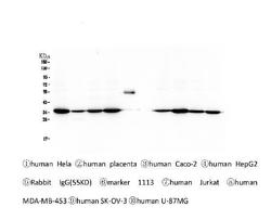
- Experimental details
- Western blot analysis of Emerin using anti-Emerin antibody (M00714). Electrophoresis was performed on a 5-20% SDS-PAGE gel at 70V (Stacking gel) / 90V (Resolving gel) for 2-3 hours. The sample well of each lane was loaded with 50ug of sample under reducing conditions. Lane 1: human Hela whole cell lysates, Lane 2: human placenta tissue lysates, Lane 3: human Caco-2 whole cell lysates, Lane 4: human HepG2 whole cell lysates,Lane 5: Rabbit IgG, Lane 6: Marker 1113, Lane 7: human Jurkat whole cell lysates. Lane 8: human MDA-MB-453 whole cell lysates, Lane 9: human SK-OV-3 whole cell lysates, Lane 10: human SW620 whole cell lysates. After Electrophoresis, proteins were transferred to a Nitrocellulose membrane at 150mA for 50-90 minutes. Blocked the membrane with 5% Non-fat Milk/ TBS for 1.5 hour at RT. The membrane was incubated with mouse anti-Emerin antigen affinity purified monoclonal antibody (Catalog # M00714) at 0.5 μg/mL overnight at 4°C, then washed with TBS-0.1%Tween 3 times with 5 minutes each and probed with a Biotin Conjugated goat anti-mouse IgG secondary antibody at a dilution of 1:10000 for 1.5 hour at RT. The signal is developed using an Enhanced Chemiluminescent detection (ECL) kit (Catalog # EK1001) with Tanon 5200 system.
- Additional image