 Explore
 Validate
 Learn
Learn Western blot
 Immunocytochemistry
 Immunohistochemistry
Antibody data
- Antibody Data
- Antigen structure
- References [4]
- Comments [0]
- Validations
- Western blot [1]
- Immunocytochemistry [1]
Submit
Validation data
Reference
Comment
Report error
- Product number
- HPA000609 - Provider product page

- Provider
- Atlas Antibodies
- Proper citation
- Atlas Antibodies Cat#HPA000609, RRID:AB_1078736
- Product name
- Anti-EMD
- Antibody type
- Polyclonal
- Description
- Polyclonal Antibody against Human EMD, Gene description: emerin, Alternative Gene Names: LEMD5, STA, Validated applications: WB, IHC, ICC, Uniprot ID: P50402, Storage: Store at +4°C for short term storage. Long time storage is recommended at -20°C.
- Reactivity
- Human, Mouse, Rat
- Host
- Rabbit
- Conjugate
- Unconjugated
- Isotype
- IgG
- Vial size
- 100 µl
- Concentration
- 0.2 mg/ml
- Storage
- Store at +4°C for short term storage. Long time storage is recommended at -20°C.
- Handling
- The antibody solution should be gently mixed before use.
Submitted references Endothelial progerin expression causes cardiovascular pathology through an impaired mechanoresponse.
Spindle associated membrane protein 1 (Samp1) is required for the differentiation of muscle cells.
Immunofluorescence and fluorescent-protein tagging show high correlation for protein localization in mammalian cells
Impact of genomic stability on protein expression in endometrioid endometrial cancer
Osmanagic-Myers S, Kiss A, Manakanatas C, Hamza O, Sedlmayer F, Szabo PL, Fischer I, Fichtinger P, Podesser BK, Eriksson M, Foisner R
The Journal of clinical investigation 2019 Feb 1;129(2):531-545
The Journal of clinical investigation 2019 Feb 1;129(2):531-545
Spindle associated membrane protein 1 (Samp1) is required for the differentiation of muscle cells.
Jafferali MH, Figueroa RA, Hasan M, Hallberg E
Scientific reports 2017 Nov 30;7(1):16655
Scientific reports 2017 Nov 30;7(1):16655
Immunofluorescence and fluorescent-protein tagging show high correlation for protein localization in mammalian cells
Stadler C, Rexhepaj E, Singan V, Murphy R, Pepperkok R, Uhlén M, Simpson J, Lundberg E
Nature Methods 2013;10(4):315-323
Nature Methods 2013;10(4):315-323
Impact of genomic stability on protein expression in endometrioid endometrial cancer
Lomnytska M, Becker S, Gemoll T, Lundgren C, Habermann J, Olsson A, Bodin I, Engström U, Hellman U, Hellman K, Hellström A, Andersson S, Mints M, Auer G
British Journal of Cancer 2012;106(7):1297-1305
British Journal of Cancer 2012;106(7):1297-1305
No comments: Submit comment
Enhanced validation
- Submitted by
- Atlas Antibodies (provider)
- Enhanced method
- Genetic validation
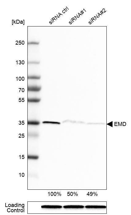
- Main image

- Experimental details
- Western blot analysis in RT-4 cells transfected with control siRNA, target specific siRNA probe #1 and #2, using Anti-EMD antibody. Remaining relative intensity is presented. Loading control: Anti-GAPDH.
- Sample type
- Human
- Protocol
- Protocol
Supportive validation
- Submitted by
- Atlas Antibodies (provider)
- Main image
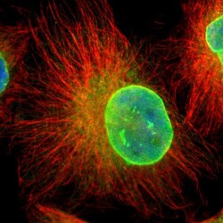
- Experimental details
- Immunofluorescent staining of human cell line U-251 MG shows localization to nuclear membrane & endoplasmic reticulum.
- Sample type
- Human
