Explore
 Validate
 Learn
Learn Western blot
 Immunocytochemistry
Antibody data
- Antibody Data
- Antigen structure
- References [2]
- Comments [0]
- Validations
- Immunocytochemistry [3]
- Immunohistochemistry [1]
Submit
Validation data
Reference
Comment
Report error
- Product number
- PA5-29731 - Provider product page

- Provider
- Invitrogen Antibodies
- Product name
- Emerin Polyclonal Antibody
- Antibody type
- Polyclonal
- Antigen
- Recombinant full-length protein
- Description
- Recommended positive controls: HeLa nucleus. Store product as a concentrated solution. Centrifuge briefly prior to opening the vial.
- Reactivity
- Human
- Host
- Rabbit
- Isotype
- IgG
- Vial size
- 100 μL
- Concentration
- 1 mg/mL
- Storage
- Store at 4°C short term. For long term storage, store at -20°C, avoiding freeze/thaw cycles.
Submitted references Combined loss of LAP1B and LAP1C results in an early onset multisystemic nuclear envelopathy.
Topokaryotyping demonstrates single cell variability and stress dependent variations in nuclear envelope associated domains.
Fichtman B, Zagairy F, Biran N, Barsheshet Y, Chervinsky E, Ben Neriah Z, Shaag A, Assa M, Elpeleg O, Harel A, Spiegel R
Nature communications 2019 Feb 5;10(1):605
Nature communications 2019 Feb 5;10(1):605
Topokaryotyping demonstrates single cell variability and stress dependent variations in nuclear envelope associated domains.
Jurisic A, Robin C, Tarlykov P, Siggens L, Schoell B, Jauch A, Ekwall K, Sørensen CS, Lipinski M, Shoaib M, Ogryzko V
Nucleic acids research 2018 Dec 14;46(22):e135
Nucleic acids research 2018 Dec 14;46(22):e135
No comments: Submit comment
Supportive validation
- Submitted by
- Invitrogen Antibodies (provider)
- Main image

- Experimental details
- Immunofluorescent analysis of Emerin in paraformaldehyde-fixed HeLa cells using an Emerin polyclonal antibody (Product # PA5-29731) at a 1:500 dilution.
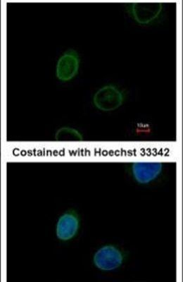
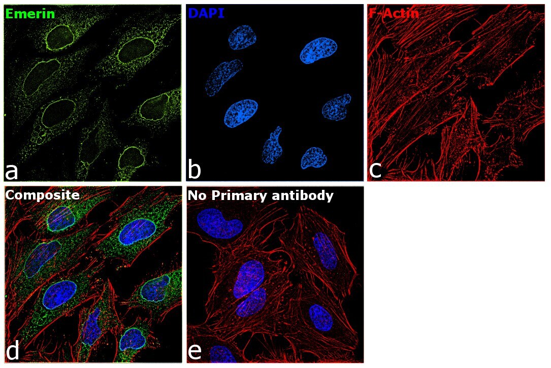
- Submitted by
- Invitrogen Antibodies (provider)
- Main image
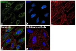
- Experimental details
- Immunofluorescence analysis of Emerin was performed using 70% confluent log phase HeLa cells. The cells were fixed with 4% paraformaldehyde for 10 minutes, permeabilized with 0.1% Triton™ X-100 for 15 minutes, and blocked with 2% BSA for 1 hour at room temperature. The cells were labeled with Emerin Polyclonal Antibody (Product # PA5-29731) at 1:250 dilution in 0.1% BSA, incubated at 4 degree Celsius overnight and then labeled with Donkey anti-Rabbit IgG (H+L) Highly Cross-Adsorbed Secondary Antibody, Alexa Fluor Plus 488 (Product # A32790) at a dilution of 1:2000 for 45 minutes at room temperature (Panel a: green). Nuclei (Panel b: blue) were stained with SlowFade® Gold Antifade Mountant with DAPI (Product # S36938). F-actin (Panel c: red) was stained with Rhodamine Phalloidin (Product # R415, 1:300). Panel d represents the merged image showing nuclear membrane and endoplasmic reticulum localization. Panel e represents control cells with no primary antibody to assess background. The images were captured at 60X magnification.
Supportive validation
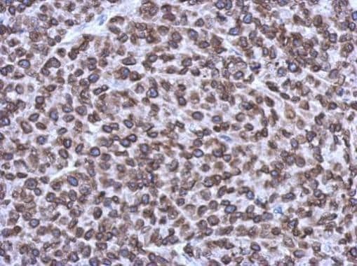
- Submitted by
- Invitrogen Antibodies (provider)
- Main image

- Experimental details
- Immunohistochemical analysis of paraffin-embedded Hela xenograft, using Emerin (Product # PA5-29731) antibody at 1:1,000 dilution. Antigen Retrieval: EDTA based buffer, pH 8.0, 15 min.