Explore
 Validate
 Learn
Learn Western blot
 Immunocytochemistry
Antibody data
- Antibody Data
- Antigen structure
- References [3]
- Comments [0]
- Validations
- Western blot [1]
Submit
Validation data
Reference
Comment
Report error
- Product number
- MA1089 - Provider product page

- Provider
- Boster Biological Technology
- Product name
- Anti-Smad4(DPC4) Antibody (monoclonal)
- Antibody type
- Monoclonal
- Antigen
- Recombinant human Smad4(DPC4).
- Description
- Ascites
- Reactivity
- Human
- Vial size
- 100μg/vial
- Concentration
- Lyophilized
- Storage
- At -20°C for one year. After reconstitution, at 4°C for one month. It can also be aliquotted and stored frozen at -20°C for a longer time. Avoid repeated freezing and thawing.
- Handling
- 1.2% sodium acetate or neutral PBS. If 1ml of PBS is used, the antibody concentration will be 100μg/ml.
Submitted references Identification of an activation-related protein in B cells in the ABO incompatible condition.
Effect of Kuijie Granule on the Expression of TGF-β/Smads Signaling Pathway in Patients with Ulcerative Colitis.
Role of transforming growth factor-beta signaling pathway in pathogenesis of benign biliary stricture.
Cao J, Chen C, Liu L, Zhang Y, Zhou H, Xiao J, Wang Y
Experimental and therapeutic medicine 2020 Jan;19(1):741-747
Experimental and therapeutic medicine 2020 Jan;19(1):741-747
Effect of Kuijie Granule on the Expression of TGF-β/Smads Signaling Pathway in Patients with Ulcerative Colitis.
Xu X, Xu C, Saud SM, Lu X, Liu L, Fang L, Zhang X, Hu J, Li W
Evidence-based complementary and alternative medicine : eCAM 2016;2016:2601830
Evidence-based complementary and alternative medicine : eCAM 2016;2016:2601830
Role of transforming growth factor-beta signaling pathway in pathogenesis of benign biliary stricture.
Geng ZM, Zheng JB, Zhang XX, Tao J, Wang L
World journal of gastroenterology 2008 Aug 21;14(31):4949-54
World journal of gastroenterology 2008 Aug 21;14(31):4949-54
No comments: Submit comment
Supportive validation
- Submitted by
- Boster Biological Technology (provider)
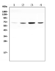
- Main image
- Experimental details
- Western blot analysis of SMAD4 using anti-SMAD4 antibody (MA1089). Electrophoresis was performed on a 5-20% SDS-PAGE gel at 70V (Stacking gel) / 90V (Resolving gel) for 2-3 hours. The sample well of each lane was loaded with 50ug of sample under reducing conditions. Lane 1: human HepG2 whole cell lysate, Lane 2: human HEK293 whole cell lysate, Lane 3: human COLO-320 whole cell lysate, Lane 4: human THP-1 whole cell lysate. After Electrophoresis, proteins were transferred to a Nitrocellulose membrane at 150mA for 50-90 minutes. Blocked the membrane with 5% Non-fat Milk/ TBS for 1.5 hour at RT. The membrane was incubated with mouse anti-SMAD4 antigen affinity purified monoclonal antibody (Catalog # MA1089) at 0.5 μg/mL overnight at 4°C, then washed with TBS-0.1%Tween 3 times with 5 minutes each and probed with a goat anti-mouse IgG-HRP secondary antibody at a dilution of 1:10000 for 1.5 hour at RT. The signal is developed using an Enhanced Chemiluminescent detection (ECL) kit (Catalog # EK1001) with Tanon 5200 system. A specific band was detected for SMAD4 at approximately 70KD. The expected band size for SMAD4 is at 60KD.
- Additional image