 Explore
 Validate
 Learn
Learn Western blot
 ELISA
ELISAAntibody data
- Antibody Data
- Antigen structure
- References [2]
- Comments [0]
- Validations
- Western blot [1]
- Immunocytochemistry [1]
- Immunohistochemistry [1]
Submit
Validation data
Reference
Comment
Report error
- Product number
- MAB623-100 - Provider product page

- Provider
- R&D Systems
- Product name
- Human CD25/IL-2R alpha Antibody
- Antibody type
- Monoclonal
- Description
- Protein A or G purified from ascites. Detects human CD25/IL-2 R alpha in ELISAs and Western blots. In Western blots, detects recombinant rat CD25/IL-2 R alpha but no cross-reactivity with recombinant mouse CD25/IL-2 R alpha . In sandwich immunoassays, this antibody does not cross-react or interfere with recombinant human (rh) IL-1 RI, rhIL-1 RII, rhIL-2 R beta , rhIL-3 R alpha , rhIL-4 R, rhIL-5 R alpha , rhIL-5 R beta , rhIL-6 R, or rhgp130.
- Reactivity
- Human
- Host
- Mouse
- Conjugate
- Unconjugated
- Antigen sequence
P01589- Isotype
- IgG
- Antibody clone number
- 24204
- Vial size
- 100 ug
- Storage
- Use a manual defrost freezer and avoid repeated freeze-thaw cycles. 12 months from date of receipt, -20 to -70 °C as supplied. 1 month, 2 to 8 °C under sterile conditions after reconstitution. 6 months, -20 to -70 °C under sterile conditions after reconstitution.
Submitted references Noninvasive detection of acute and chronic injuries in human renal transplant by elevation of multiple cytokines/chemokines in urine.
Imbalance in the peritoneal levels of interleukin 1 and its decoy inhibitory receptor type II in endometriosis women with infertility and pelvic pain.
Hu H, Kwun J, Aizenstein BD, Knechtle SJ
Transplantation 2009 Jun 27;87(12):1814-20
Transplantation 2009 Jun 27;87(12):1814-20
Imbalance in the peritoneal levels of interleukin 1 and its decoy inhibitory receptor type II in endometriosis women with infertility and pelvic pain.
Akoum A, Al-Akoum M, Lemay A, Maheux R, Leboeuf M
Fertility and sterility 2008 Jun;89(6):1618-24
Fertility and sterility 2008 Jun;89(6):1618-24
No comments: Submit comment
Supportive validation
- Submitted by
- R&D Systems (provider)
- Main image
- Experimental details
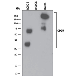
- Detection of Recombinant Human and Rat CD25/IL-2 R alpha by Western Blot. Western blot shows 25 ng of Recombinant Human CD25/IL-2 R alpha (Catalog # 223-2A), Recombinant Mouse CD25/IL-2 R alpha (Catalog # 2438-RM) and Recombinant Rat CD25/IL-2 R alpha Fc Chimera (Catalog # 5156-RM). PVDF Membrane was probed with 1 µg/mL of Mouse Anti-Human CD25/IL-2 R alpha Monoclonal Antibody (Catalog # MAB623) followed by HRP-conjugated Anti-Mouse IgG Secondary Antibody (Catalog # HAF018). Specific bands were detected for CD25/IL-2 R alpha at approximately 30-250 kDa (as indicated). This experiment was conducted under non-reducing conditions and using Immunoblot Buffer Group 3.

Supportive validation
- Submitted by
- R&D Systems (provider)
- Main image
- Experimental details
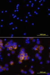
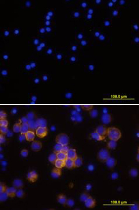
- CD25/IL-2 R alpha in Human PBMCs. CD25/IL-2 R alpha was detected in immersion fixed human peripheral blood mononuclear cells (PBMCs) stimulated with PHA using Mouse Anti-Human CD25/IL-2 R alpha Monoclonal Antibody (Catalog # MAB623) at 10 µg/mL for 3 hours at room temperature. Cells were stained using the NorthernLights™ 557-conjugated Anti-Mouse IgG Secondary Antibody (yellow; Catalog # NL007) and counterstained with DAPI (blue). Upper panel shows a lack of labeling if primary antibodies are omitted and tissue is stained only with secondary antibody followed by incubation with detection reagents. View our protocol for Fluorescent ICC Staining of Non-adherent Cells.
Supportive validation
- Submitted by
- R&D Systems (provider)
- Main image
- Experimental details
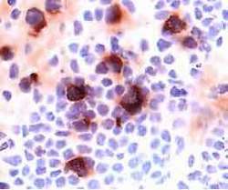
- CD25/IL-2 R alpha in Human Lymph Node. CD25/IL-2 R alpha was detected in immersion fixed frozen sections of human lymph node using 15 µg/mL Mouse Anti-Human CD25/IL-2 R alpha Monoclonal Antibody (Catalog # MAB623) overnight at 4 °C. Tissue was stained (red) and counterstained with hematoxylin (blue). View our protocol for Chromogenic IHC Staining of Frozen Tissue Sections.
