Explore
 Validate
 Learn
Learn Flow cytometry
Antibody data
- Antibody Data
- Antigen structure
- References [8]
- Comments [0]
- Validations
- Flow cytometry [1]
- Other assay [3]
Submit
Validation data
Reference
Comment
Report error
- Product number
- 11-1379-42 - Provider product page

- Provider
- Invitrogen Antibodies
- Product name
- CD137 (4-1BB) Monoclonal Antibody (4B4 (4B4-1)), FITC, eBioscience™
- Antibody type
- Monoclonal
- Antigen
- Other
- Description
- Description: This 4B4 (4B4-1) monoclonal antibody reacts with human CD137 (also known as 4-1BB or TNFRSF9), which is an inducible member of the TNFR family of costimulatory molecules expressed on T cells, natural killer cells, dendritic cells, granulocytes, and mast cells. Involved in recruiting TNFR-associated factors (TRAF) 1 and 2, CD137 signaling plays a role in T cell activation, maintaining the survival of activated and CD8 memory T cells, as well as suppressing myelopoiesis and dendritic cell development. Stimulation of this receptor has also been shown to promote expansion of CD4+CD25+ T regulatory cells \i{ex vivo\i}. The ligand for CD137, 4-1BBL, is found on activated macrophages, mature B cells, hematopoietic stem cells, and myeloid progenitor cells. Applications Reported: This 4B4 (4B4-1) antibody has been reported for use in flow cytometric analysis. Applications Tested: This 4B4 (4B4-1) antibody has been pre-titrated and tested by flow cytometric analysis on Con A-stimulated normal human peripheral blood cells. This can be used at 5 µL (0.06 µg) per test. A test is defined as the amount (µg) of antibody that will stain a cell sample in a final volume of 100 µL. Cell number should be determined empirically but can range from 10^5 to 10^8 cells/test. Excitation: 488 nm; Emission: 520 nm; Laser: Blue Laser. Filtration: 0.2 µm post-manufacturing filtered.
- Reactivity
- Human
- Host
- Mouse
- Conjugate
- Green dye
- Isotype
- IgG
- Antibody clone number
- 4B4 (4B4-1)
- Vial size
- 100 Tests
- Concentration
- 5 µL/Test
- Storage
- 4° C, store in dark, DO NOT FREEZE!
Submitted references Neoantigens retention in patient derived xenograft models mediates autologous T cells activation in ovarian cancer.
A transcriptionally and functionally distinct PD-1(+) CD8(+) T cell pool with predictive potential in non-small-cell lung cancer treated with PD-1 blockade.
Antroquinonol Exerts Immunosuppressive Effect on CD8(+) T Cell Proliferation and Activation to Resist Depigmentation Induced by H(2)O(2).
Surrogate in vitro activation of innate immunity synergizes with interleukin-7 to unleash rapid antigen-driven outgrowth of CD4+ and CD8+ human peripheral blood T-cells naturally recognizing MUC1, HER2/neu and other tumor-associated antigens.
ERK-dependent Bim modulation downstream of the 4-1BB-TRAF1 signaling axis is a critical mediator of CD8 T cell survival in vivo.
Identification of regulatory functions for 4-1BB and 4-1BBL in myelopoiesis and the development of dendritic cells.
Ex vivo expansion of CD4+CD25+FoxP3+ T regulatory cells based on synergy between IL-2 and 4-1BB signaling.
4-1BB is expressed on CD45RAhiROhi transitional T cell in humans.
Want MY, Konstorum A, Huang RY, Jain V, Matsueda S, Tsuji T, Lugade A, Odunsi K, Koya R, Battaglia S
Oncoimmunology 2019;8(6):e1586042
Oncoimmunology 2019;8(6):e1586042
A transcriptionally and functionally distinct PD-1(+) CD8(+) T cell pool with predictive potential in non-small-cell lung cancer treated with PD-1 blockade.
Thommen DS, Koelzer VH, Herzig P, Roller A, Trefny M, Dimeloe S, Kiialainen A, Hanhart J, Schill C, Hess C, Savic Prince S, Wiese M, Lardinois D, Ho PC, Klein C, Karanikas V, Mertz KD, Schumacher TN, Zippelius A
Nature medicine 2018 Jul;24(7):994-1004
Nature medicine 2018 Jul;24(7):994-1004
Antroquinonol Exerts Immunosuppressive Effect on CD8(+) T Cell Proliferation and Activation to Resist Depigmentation Induced by H(2)O(2).
Guan C, Li Q, Song X, Xu W, Li L, Xu A
Oxidative medicine and cellular longevity 2017;2017:9303054
Oxidative medicine and cellular longevity 2017;2017:9303054
Surrogate in vitro activation of innate immunity synergizes with interleukin-7 to unleash rapid antigen-driven outgrowth of CD4+ and CD8+ human peripheral blood T-cells naturally recognizing MUC1, HER2/neu and other tumor-associated antigens.
Pathangey LB, McCurry DB, Gendler SJ, Dominguez AL, Gorman JE, Pathangey G, Mihalik LA, Dang Y, Disis ML, Cohen PA
Oncotarget 2017 Feb 14;8(7):10785-10808
Oncotarget 2017 Feb 14;8(7):10785-10808
ERK-dependent Bim modulation downstream of the 4-1BB-TRAF1 signaling axis is a critical mediator of CD8 T cell survival in vivo.
Sabbagh L, Pulle G, Liu Y, Tsitsikov EN, Watts TH
Journal of immunology (Baltimore, Md. : 1950) 2008 Jun 15;180(12):8093-101
Journal of immunology (Baltimore, Md. : 1950) 2008 Jun 15;180(12):8093-101
Identification of regulatory functions for 4-1BB and 4-1BBL in myelopoiesis and the development of dendritic cells.
Lee SW, Park Y, So T, Kwon BS, Cheroutre H, Mittler RS, Croft M
Nature immunology 2008 Aug;9(8):917-26
Nature immunology 2008 Aug;9(8):917-26
Ex vivo expansion of CD4+CD25+FoxP3+ T regulatory cells based on synergy between IL-2 and 4-1BB signaling.
Elpek KG, Yolcu ES, Franke DD, Lacelle C, Schabowsky RH, Shirwan H
Journal of immunology (Baltimore, Md. : 1950) 2007 Dec 1;179(11):7295-304
Journal of immunology (Baltimore, Md. : 1950) 2007 Dec 1;179(11):7295-304
4-1BB is expressed on CD45RAhiROhi transitional T cell in humans.
Garni-Wagner BA, Lee ZH, Kim YJ, Wilde C, Kang CY, Kwon BS
Cellular immunology 1996 Apr 10;169(1):91-8
Cellular immunology 1996 Apr 10;169(1):91-8
No comments: Submit comment
Supportive validation
- Submitted by
- Invitrogen Antibodies (provider)
- Main image

- Experimental details
- Staining of ConA-stimulated human peripheral blood cells with Anti-Human CD69 APC (Product # 17-0699-42) and Mouse IgG1 kappa Isotype Control FITC (Product # 11-4714-42) (left) or Anti-Human CD137 (4-1BB) FITC (right). Total viable cells were used for analysis.
- Conjugate
- Green dye

Supportive validation
- Submitted by
- Invitrogen Antibodies (provider)
- Main image

- Experimental details
- NULL
- Conjugate
- Green dye

- Submitted by
- Invitrogen Antibodies (provider)
- Main image
- Experimental details
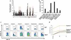
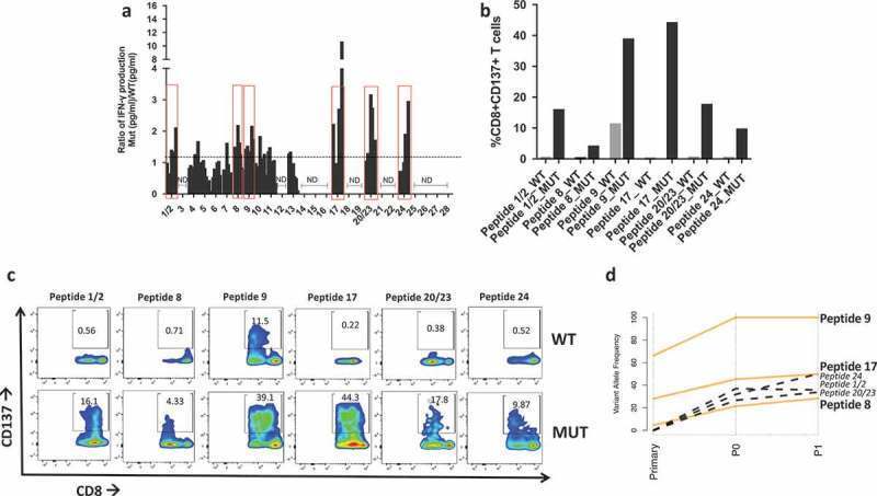
- 10.1080/2162402X.2019.1586042-F0004 Figure 4. Neoantigen recognition by autologous T cells in-vitro . a) Ratio of IFN-gamma production in patient's T cells at different time points upon coculture with mutated (MUT) or WT peptide-stimulated autologous APCs. Y axis indicates the ratio IFN-gamma Mut(pg/ml)/IFN-gamma WT(pg/ml) measured via ELISA. Peptides with a ratio > 1 are highlighted in red boxes and utilized for subsequent experiments. ND = no IFN-gamma production detected. b) Flow cytometry analysis of CD137 + T cells expressed as percent of activated CD8 + T cells after overnight stimulation with 10 um mutated (MUT) or WT peptides. c) Flow plots of CD137 + /CD8 + T cells in response to coculture with APC pulsed with mutated (MUT) or WT peptides. d) Line plot showing the changes in variant allele frequency of the six neoantigenic mutations common across the primary, P0 and P1 tumors. Orange lines represent mutations present in all samples, black dotted mutations indicate those present in P0 and P1 but absent (VAF = 0) in the primary tumor.
- Conjugate
- Green dye
- Submitted by
- Invitrogen Antibodies (provider)
- Main image
- Experimental details
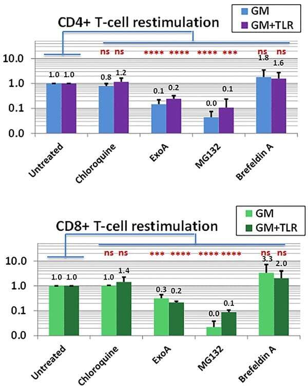
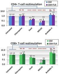
- Figure 6 Exposure to R848 and LPS is a critical determinant of CD8+ T-cell sensitization A . left panel. IL-12 and IL-23 production during first two days of culture are both markedly enhanced by dual stimulation with R848 and LPS. Methods as in Figure 1B . Note that cytokine production is shown in log scale. This graphing of ELISA results shows an averaging of 4 biological replicates. A . right panel, exposure to R848+LPS enhances frequency and/or density of co-stimulatory ligands and receptors on PBMC CD33+ constituents. Methods as in Figure 1B ., with FACS analyses performed after 48h in culture. Frequency of each molecule of interest in the CD33+ subpopulation was determined and averaged for 4 biological replicates; in addition, fold-increase in cell surface staining density relative to isotype control (averaged Mean Fluorescent Index) was determined. Frequency of expression is shown graphically, whereas for MFI only tabulated p values are shown. Exposure to R848+LPS significantly increased both the frequency and MFI of CD70, 4-1BB ligand, and B7.1 (CD80) (shown on graph). In addition, although frequency of CD40 expression was already nearly uniform without R848+LPS exposure, R848+LPS significantly increased the MFI for CD40. Two-tailed p values are shown, based on a paired Student t -test comparing with and without R848+LPS exposure within each experimental run. B, left panel, dual exposure to R848+LPS significantly enhances Ag-specific CD4+ and CD8+ propagation. Cultures
- Conjugate
- Green dye