Explore
 Validate
 Learn
Learn Flow cytometry
Antibody data
- Antibody Data
- Antigen structure
- References [2]
- Comments [0]
- Validations
- Flow cytometry [1]
- Other assay [2]
Submit
Validation data
Reference
Comment
Report error
- Product number
- 46-1379-41 - Provider product page

- Provider
- Invitrogen Antibodies
- Product name
- CD137 (4-1BB) Monoclonal Antibody (4B4 (4B4-1)), PerCP-eFluor™ 710, eBioscience™
- Antibody type
- Monoclonal
- Antigen
- Other
- Description
- Description: This 4B4 (4B4-1) monoclonal antibody reacts with human CD137 (also known as 4-1BB or TNFRSF9), which is an inducible member of the TNFR family of costimulatory molecules expressed on T cells, natural killer cells, dendritic cells, granulocytes, and mast cells. Involved in recruiting TNFR-associated factors (TRAF) 1 and 2, CD137 signaling plays a role in T cell activation, maintaining the survival of activated and CD8 memory T cells, as well as suppressing myelopoiesis and dendritic cell development. Stimulation of this receptor has also been shown to promote expansion of CD4+CD25+ T regulatory cells \i{ex vivo\i}. The ligand for CD137, 4-1BBL, is found on activated macrophages, mature B cells, hematopoietic stem cells, and myeloid progenitor cells. Applications Reported: This 4B4 (4B4-1) antibody has been reported for use in flow cytometric analysis. Applications Tested: This 4B4 (4B4-1) antibody has been pre-titrated and tested by flow cytometric analysis of stimulated normal human peripheral blood cells. This can be used at 5 µL (0.125 µg) per test. A test is defined as the amount (µg) of antibody that will stain a cell sample in a final volume of 100 µL. Cell number should be determined empirically but can range from 10^5 to 10^8 cells/test. PerCP-eFluor® 710 emits at 710 nm and is excited with the blue laser (488 nm); it can be used in place of PerCP-Cyanine5.5. We recommend using a 710/50 bandpass filter, however, the 695/40 bandpass filter is an acceptable alternative. Please make sure that your instrument is capable of detecting this fluorochrome. Fixation: Samples can be stored in IC Fixation Buffer (Product # 00-822-49) (100 µL cell sample + 100 µL IC Fixation Buffer) or 1-step Fix/Lyse Solution (Product # 00-5333-54) for up to 3 days in the dark at 4°C with minimal impact on brightness and FRET efficiency/compensation. Some generalizations regarding fluorophore performance after fixation can be made, but clone specific performance should be determined empirically. Excitation: 488 nm; Emission: 710 nm; Laser: Blue Laser. Filtration: 0.2 µm post-manufacturing filtered.
- Reactivity
- Human
- Host
- Mouse
- Isotype
- IgG
- Antibody clone number
- 4B4 (4B4-1)
- Vial size
- 25 Tests
- Concentration
- 5 µL/Test
- Storage
- 4° C, store in dark, DO NOT FREEZE!
Submitted references Neoantigens retention in patient derived xenograft models mediates autologous T cells activation in ovarian cancer.
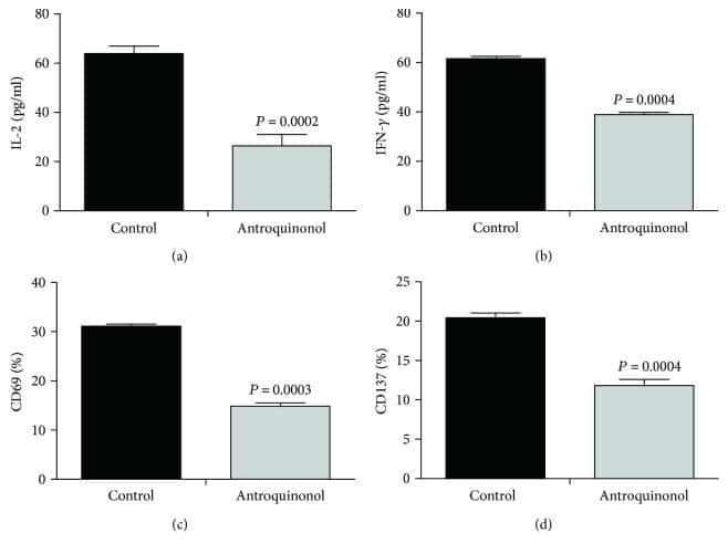
Antroquinonol Exerts Immunosuppressive Effect on CD8(+) T Cell Proliferation and Activation to Resist Depigmentation Induced by H(2)O(2).
Want MY, Konstorum A, Huang RY, Jain V, Matsueda S, Tsuji T, Lugade A, Odunsi K, Koya R, Battaglia S
Oncoimmunology 2019;8(6):e1586042
Oncoimmunology 2019;8(6):e1586042
Antroquinonol Exerts Immunosuppressive Effect on CD8(+) T Cell Proliferation and Activation to Resist Depigmentation Induced by H(2)O(2).
Guan C, Li Q, Song X, Xu W, Li L, Xu A
Oxidative medicine and cellular longevity 2017;2017:9303054
Oxidative medicine and cellular longevity 2017;2017:9303054
No comments: Submit comment
Supportive validation
- Submitted by
- Invitrogen Antibodies (provider)
- Main image
- Experimental details
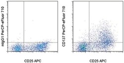
- Staining of 3-day Con A-stimulated normal human peripheral blood cells with Anti-Human CD25 APC (Product # 17-0257-42) and Mouse IgG1 K Isotype Control PerCP-eFluor® 710 (Product # 46-4714-82) (left) or Anti-Human CD137 (4-1BB) PerCP-eFluor® 710 (right). Viable cells, as determined by Fixable Viability Dye eFluor® 450 (Product # 65-0863-14), in the lymphocyte gate were used for analysis.

Supportive validation
- Submitted by
- Invitrogen Antibodies (provider)
- Main image

- Experimental details
- NULL
- Submitted by
- Invitrogen Antibodies (provider)
- Main image
- Experimental details
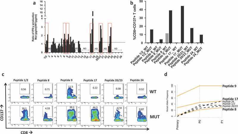
- 10.1080/2162402X.2019.1586042-F0004 Figure 4. Neoantigen recognition by autologous T cells in-vitro . a) Ratio of IFN-gamma production in patient's T cells at different time points upon coculture with mutated (MUT) or WT peptide-stimulated autologous APCs. Y axis indicates the ratio IFN-gamma Mut(pg/ml)/IFN-gamma WT(pg/ml) measured via ELISA. Peptides with a ratio > 1 are highlighted in red boxes and utilized for subsequent experiments. ND = no IFN-gamma production detected. b) Flow cytometry analysis of CD137 + T cells expressed as percent of activated CD8 + T cells after overnight stimulation with 10 um mutated (MUT) or WT peptides. c) Flow plots of CD137 + /CD8 + T cells in response to coculture with APC pulsed with mutated (MUT) or WT peptides. d) Line plot showing the changes in variant allele frequency of the six neoantigenic mutations common across the primary, P0 and P1 tumors. Orange lines represent mutations present in all samples, black dotted mutations indicate those present in P0 and P1 but absent (VAF = 0) in the primary tumor.