 Explore
 Validate
 Learn
Learn Western blot
 ELISA
ELISAAntibody data
- Antibody Data
- Antigen structure
- References [4]
- Comments [0]
- Validations
- Western blot [2]
Submit
Validation data
Reference
Comment
Report error
- Product number
- MAB972-100 - Provider product page

- Provider
- R&D Systems
- Product name
- Human EMMPRIN/CD147 Antibody
- Antibody type
- Monoclonal
- Description
- Protein A or G purified from hybridoma culture supernatant. Detects human EMMPRIN/CD147 in direct ELISAs and Western blots. In direct ELISAs and Western blots, no cross-reactivity with recombinant mouse EMMPRIN is observed.
- Reactivity
- Human
- Host
- Mouse
- Conjugate
- Unconjugated
- Antigen sequence
Q54A51- Isotype
- IgG
- Antibody clone number
- 109403
- Vial size
- 100 ug
- Storage
- Use a manual defrost freezer and avoid repeated freeze-thaw cycles. 12 months from date of receipt, -20 to -70 °C as supplied. 1 month, 2 to 8 °C under sterile conditions after reconstitution. 6 months, -20 to -70 °C under sterile conditions after reconstitution.
Submitted references The net acid extruders NHE1, NBCn1 and MCT4 promote mammary tumor growth through distinct but overlapping mechanisms.
Immunolocalization of EMMPRIN (CD147) in the human eye and detection of soluble form of EMMPRIN in ocular fluids.
Membrane type 1 matrix metalloproteinase (MT1-MMP/MMP-14) cleaves and releases a 22-kDa extracellular matrix metalloproteinase inducer (EMMPRIN) fragment from tumor cells.
Oxidized low-density lipoproteins stimulate extracellular matrix metalloproteinase Inducer (EMMPRIN) release by coronary smooth muscle cells.
Andersen AP, Samsøe-Petersen J, Oernbo EK, Boedtkjer E, Moreira JMA, Kveiborg M, Pedersen SF
International journal of cancer 2018 Jun 15;142(12):2529-2542
International journal of cancer 2018 Jun 15;142(12):2529-2542
Immunolocalization of EMMPRIN (CD147) in the human eye and detection of soluble form of EMMPRIN in ocular fluids.
Määttä M, Tervahartiala T, Kaarniranta K, Tang Y, Yan L, Tuukkanen J, Sorsa T
Current eye research 2006 Nov;31(11):917-24
Current eye research 2006 Nov;31(11):917-24
Membrane type 1 matrix metalloproteinase (MT1-MMP/MMP-14) cleaves and releases a 22-kDa extracellular matrix metalloproteinase inducer (EMMPRIN) fragment from tumor cells.
Egawa N, Koshikawa N, Tomari T, Nabeshima K, Isobe T, Seiki M
The Journal of biological chemistry 2006 Dec 8;281(49):37576-85
The Journal of biological chemistry 2006 Dec 8;281(49):37576-85
Oxidized low-density lipoproteins stimulate extracellular matrix metalloproteinase Inducer (EMMPRIN) release by coronary smooth muscle cells.
Haug C, Lenz C, Díaz F, Bachem MG
Arteriosclerosis, thrombosis, and vascular biology 2004 Oct;24(10):1823-9
Arteriosclerosis, thrombosis, and vascular biology 2004 Oct;24(10):1823-9
No comments: Submit comment
Supportive validation
- Submitted by
- R&D Systems (provider)
- Main image
- Experimental details
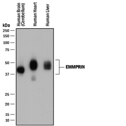
- Detection of Human EMMPRIN/CD147 by Western Blot. Western blot shows lysates of human brain (cerebellum) tissue, human heart tissue, and human liver tissue. PVDF membrane was probed with 0.2 µg/mL of Mouse Anti-Human EMMPRIN/CD147 Monoclonal Antibody (Catalog # MAB972) followed by HRP-conjugated Anti-Mouse IgG Secondary Antibody (Catalog # HAF018). A specific band was detected for EMMPRIN/CD147 at approximately 40-60 kDa (as indicated). This experiment was conducted under reducing conditions and using Immunoblot Buffer Group 1.

- Submitted by
- R&D Systems (provider)
- Main image
- Experimental details
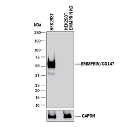
- Western Blot Shows Human EMMPRIN/CD147 Specificity by Using Knockout Cell Line. Western blot shows lysates of HEK293T human embryonic kidney parental cell line and EMMPRIN/CD147 knockout HEK293T cell line (KO). PVDF membrane was probed with 0.2 µg/mL of Mouse Anti-Human EMMPRIN/CD147 Monoclonal Antibody (Catalog # MAB972) followed by HRP-conjugated Anti-Mouse IgG Secondary Antibody (Catalog # HAF018). A specific band was detected for EMMPRIN/CD147 at approximately 51 kDa (as indicated) in the parental HEK293T cell line, but is not detectable in knockout HEK293T cell line. GAPDH (Catalog # MAB5718) is shown as a loading control. This experiment was conducted under reducing conditions and using Immunoblot Buffer Group 1.
