Explore
 Validate
 Learn
Learn Western blot
 ELISA
ELISA Immunocytochemistry
Antibody data
- Antibody Data
- Antigen structure
- References [0]
- Comments [0]
- Validations
- Immunocytochemistry [2]
- Immunohistochemistry [7]
- Flow cytometry [2]
- Other assay [1]
Submit
Validation data
Reference
Comment
Report error
- Product number
- MA5-29060 - Provider product page

- Provider
- Invitrogen Antibodies
- Product name
- CD147 Recombinant Rabbit Monoclonal Antibody (125)
- Antibody type
- Monoclonal
- Antigen
- Recombinant full-length protein
- Description
- This product is preservative free. It is recommended to add sodium azide to avoid contamination (final concentration 0.05%-0.1%). Recombinant rabbit monoclonal antibodies are produced using in vitro expression systems. The expression systems are developed by cloning in the specific antibody DNA sequences from immunoreactive rabbits. Then, individual clones are screened to select the best candidates for production. The advantages of using recombinant rabbit monoclonal antibodies include: better specificity and sensitivity, lot-to-lot consistency, animal origin-free formulations, and broader immunoreactivity to diverse targets due to larger rabbit immune repertoire. This antibody has specificity for Human CD147/Basigin/BSG.
- Reactivity
- Human
- Host
- Rabbit
- Isotype
- IgG
- Antibody clone number
- 125
- Vial size
- 100 μL
- Concentration
- 1 mg/mL
- Storage
- Store at 4°C short term. For long term storage, store at -20°C, avoiding freeze/thaw cycles.
No comments: Submit comment
Supportive validation
- Submitted by
- Invitrogen Antibodies (provider)
- Main image

- Experimental details
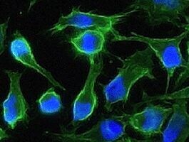
- Immunofluorescence staining of human CD147 in HeLa cells with rabbit monoclonal antibody (1:200). The image showing membrane staining of HeLa cells. CD147 Recombinant Rabbit Monoclonal Antibody (125) (Product # MA5-29060).
- Submitted by
- Invitrogen Antibodies (provider)
- Main image

- Experimental details
- Immunofluorescence staining of human CD147 in HeLa cells with rabbit monoclonal antibody (1:200). The image showing membrane staining of HeLa cells. CD147 Recombinant Rabbit Monoclonal Antibody (125) (Product # MA5-29060).

Supportive validation
- Submitted by
- Invitrogen Antibodies (provider)
- Main image
- Experimental details
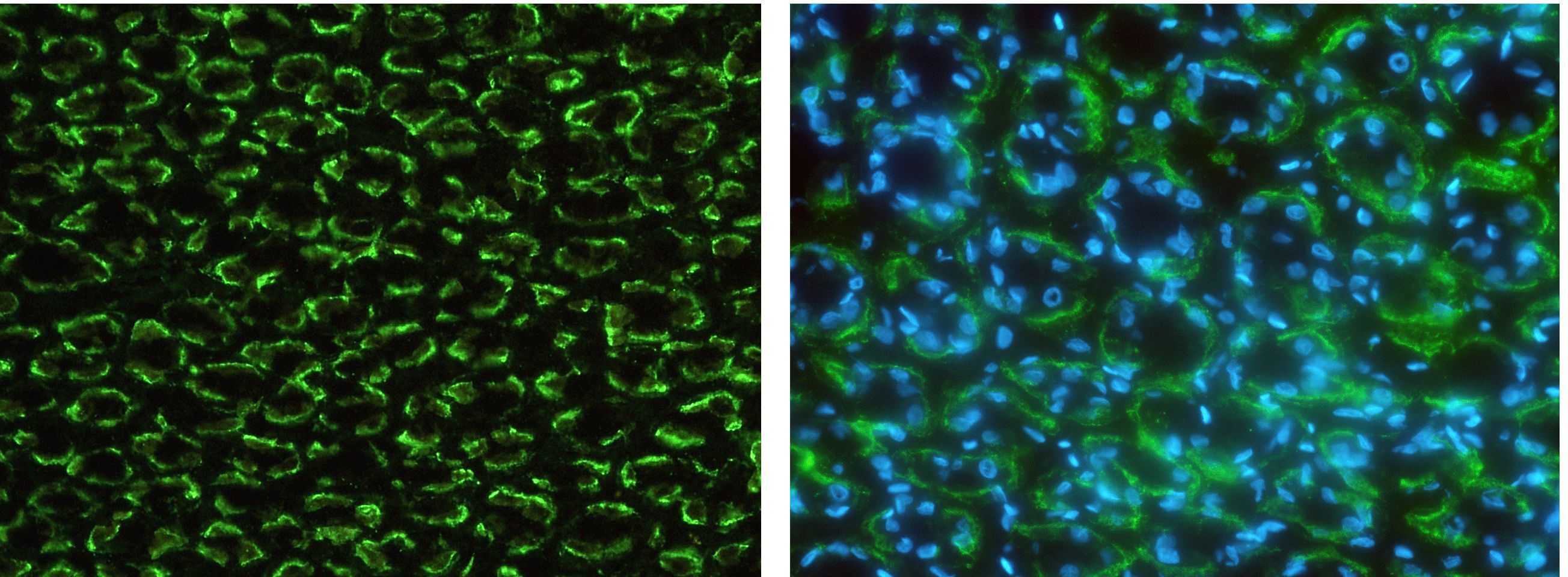
- Immunohistochemistry (Frozen) staining of CD147 in monkey stomach with CD147 Recombinant Rabbit Monoclonal Antibody (125) (Product # MA5-29060) (1:1,000, frozen section). The image showing membrane staining of gastric gland cell. Right panel: merge with DAPI.

- Submitted by
- Invitrogen Antibodies (provider)
- Main image
- Experimental details
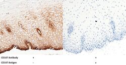
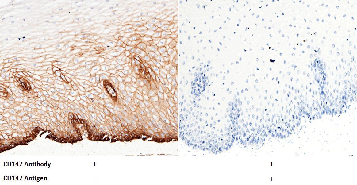
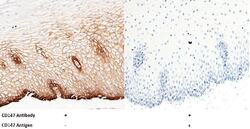
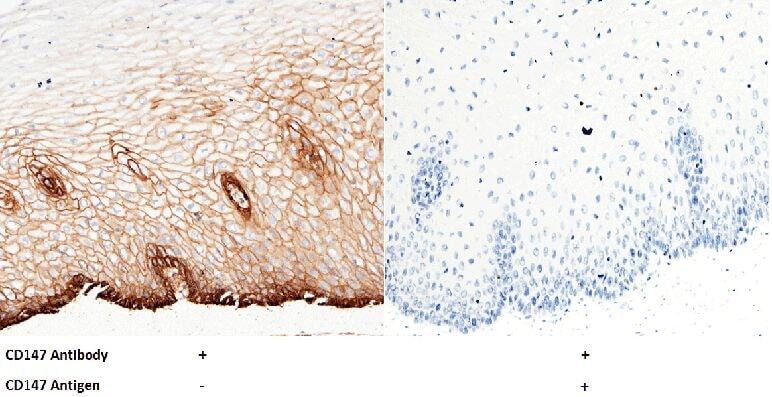
- Immunohistochemical staining of human CD147 in human esophagus with CD147 Recombinant Rabbit Monoclonal Antibody (125) (Product # MA5-29060, 1:5,000, formalin-fixed paraffin embedded sections). The image showing membrane staining of squamous epithelium cell. Left panel: tissue incubated with primary antibody. Right panel: tissue incubated with mixture of primary antibody and antigen (recombinant protein).
- Submitted by
- Invitrogen Antibodies (provider)
- Main image
- Experimental details
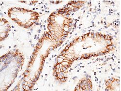
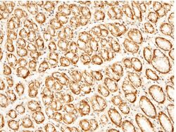
- Immunohistochemical staining of human CD147 in human gastric cancer with CD147 Recombinant Rabbit Monoclonal Antibody (125) (Product # MA5-29060, 1:5,000, formalin-fixed paraffin embedded sections). The image showing membrane staining of epithelium cell.
- Submitted by
- Invitrogen Antibodies (provider)
- Main image

- Experimental details
- Immunohistochemical staining of human CD147 in human liver with CD147 Recombinant Rabbit Monoclonal Antibody (125) (Product # MA5-29060, 1:5,000, formalin-fixed paraffin embedded sections). The image showing membrane staining of hepatocyte.
- Submitted by
- Invitrogen Antibodies (provider)
- Main image
- Experimental details
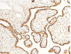
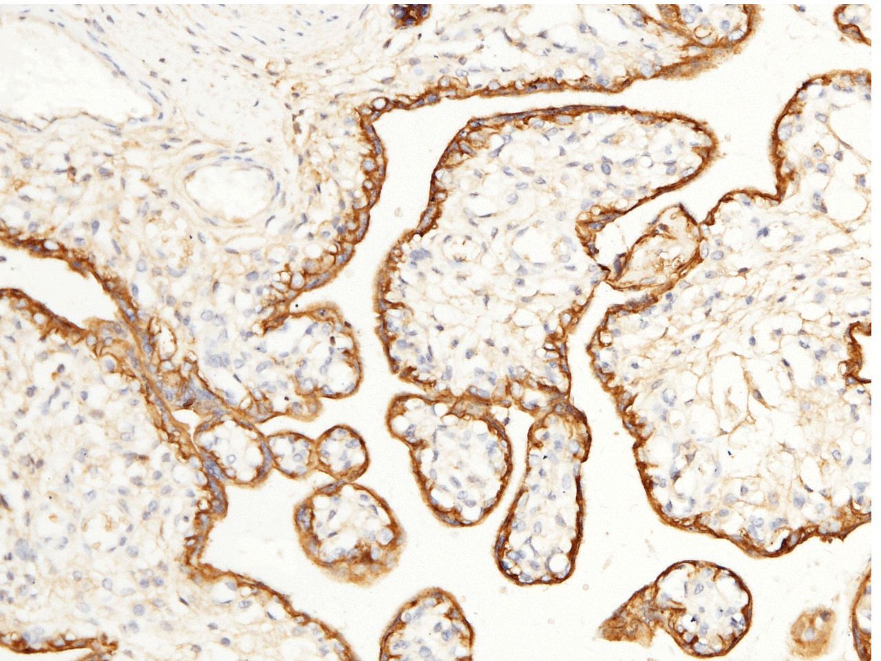
- Immunohistochemical staining of human CD147 in human placenta with CD147 Recombinant Rabbit Monoclonal Antibody (125) (Product # MA5-29060, 1:5,000, formalin-fixed paraffin embedded sections). The image showing Positive staining trophoblast.
- Submitted by
- Invitrogen Antibodies (provider)
- Main image

- Experimental details
- Immunohistochemical staining of human CD147 in human stomach with CD147 Recombinant Rabbit Monoclonal Antibody (125) (Product # MA5-29060, 1:5,000, formalin-fixed paraffin embedded sections). The image showing membrane staining of epithelium cell.
- Submitted by
- Invitrogen Antibodies (provider)
- Main image
- Experimental details
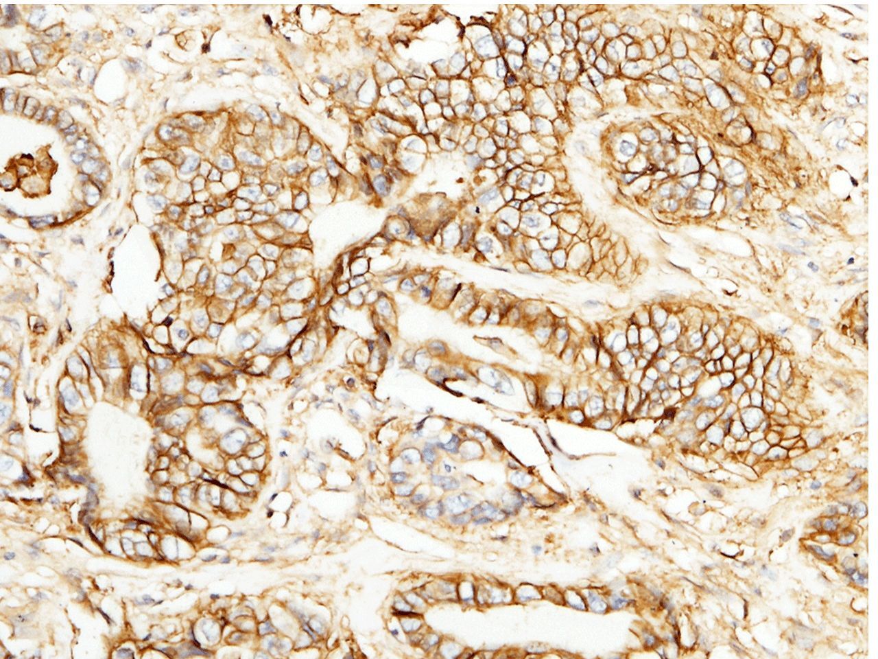
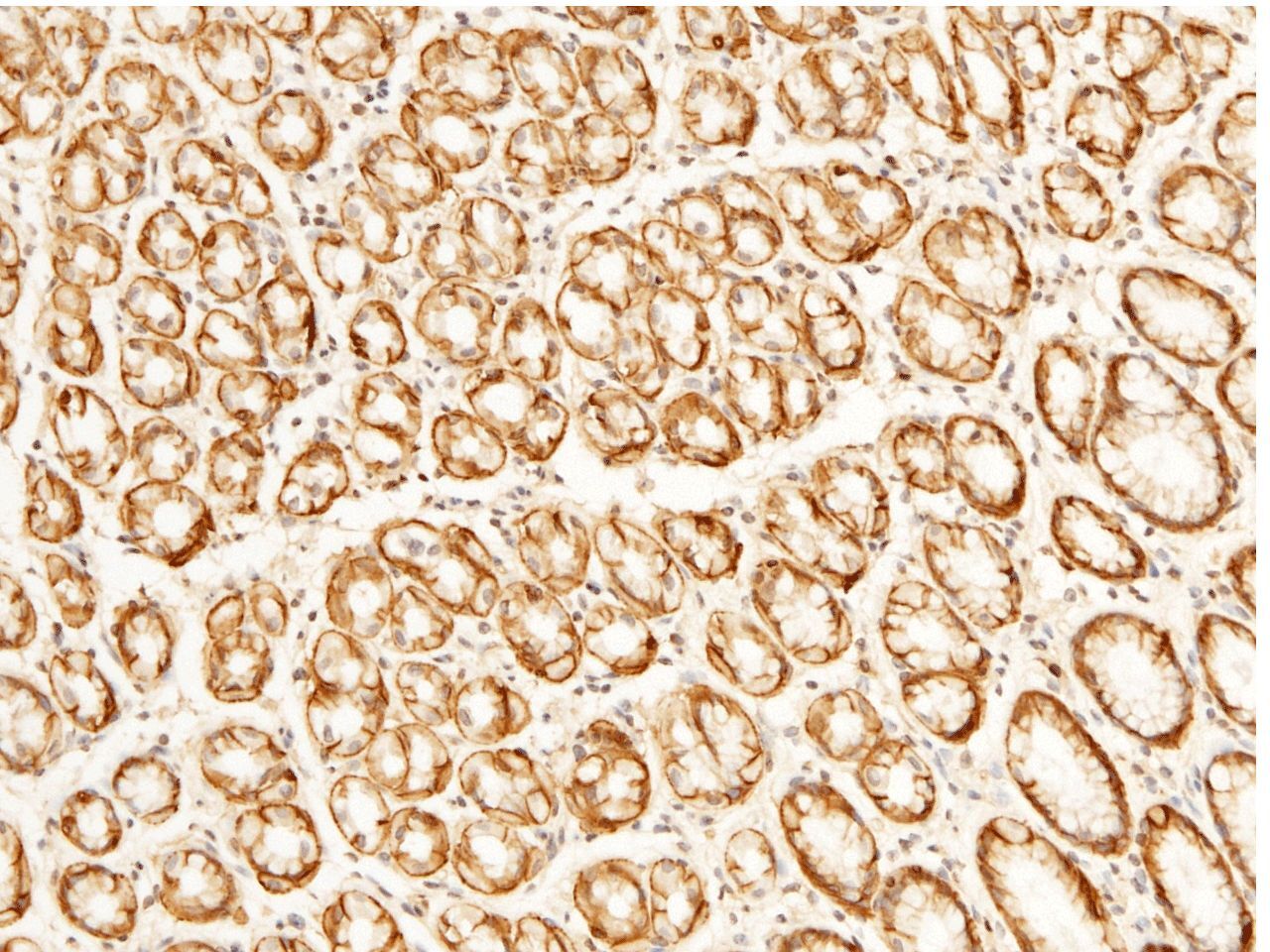
- Immunohistochemical staining of human CD147 in human rectal cancer with CD147 Recombinant Rabbit Monoclonal Antibody (125) (Product # MA5-29060, 1:5,000, formalin-fixed paraffin embedded sections). The image showing membrane staining of epithelium cell in intestinal gland.
Supportive validation
- Submitted by
- Invitrogen Antibodies (provider)
- Main image

- Experimental details
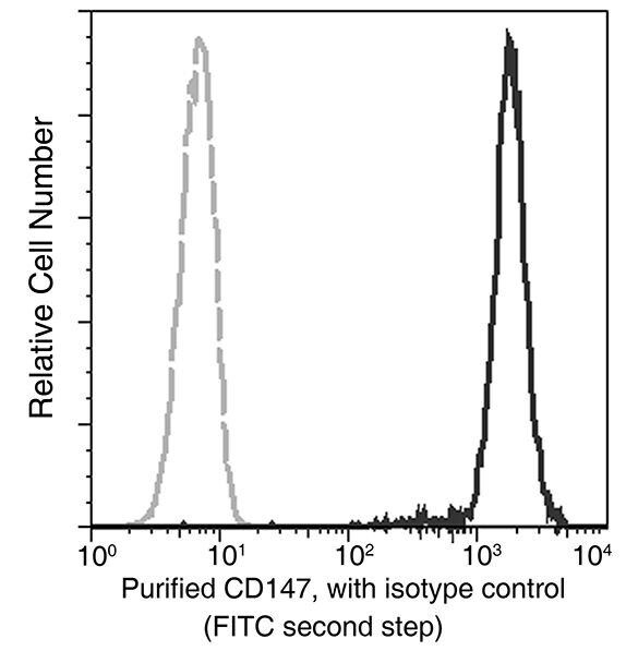
- Flow cytometry analysis of anti-CD147 reactivity on HeLa cells using CD147 monoclonal antibody (Product # MA5-29060).

- Submitted by
- Invitrogen Antibodies (provider)
- Main image

- Experimental details
- Flow cytometry analysis of anti-CD147 reactivity on HeLa cells using CD147 monoclonal antibody (Product # MA5-29060).
Supportive validation
- Submitted by
- Invitrogen Antibodies (provider)
- Main image
- Experimental details
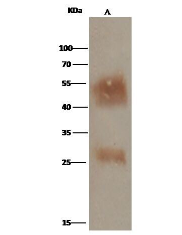
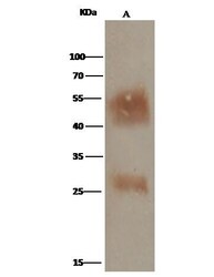
- CD147 Immunoprecipitation using: Lane A: 0.5 mg K562 Whole Cell Lysate 2 µL with CD147 Recombinant Rabbit Monoclonal Antibody (125) (Product # MA5-29060) and 15 µL of 50 % Protein G agarose. Primary antibody: CD147 Recombinant Rabbit Monoclonal Antibody (125), at 1:100 dilution. Secondary antibody: Clean-Bloto IP Detection Reagent (HRP) at 1:1,000 dilution. Developed using the DAB staining technique. Performed under reducing conditions. Predicted band size: 42 kDa. Observed band size: 50 kDa.