 Explore
 Validate
 Learn
Learn Western blot
 Immunocytochemistry
 Immunohistochemistry
Antibody data
- Antibody Data
- Antigen structure
- References [0]
- Comments [0]
- Validations
- Immunocytochemistry [3]
Submit
Validation data
Reference
Comment
Report error
- Product number
- LS-C204575 - Provider product page

- Provider
- LSBio
- Product name
- Peripherin Antibody LS-C204575
- Antibody type
- Polyclonal
- Description
- Antiserum
- Reactivity
- Human
- Host
- Rabbit
- Storage
- Store at 4°C or -20°C. Avoid freeze-thaw cycles.
No comments: Submit comment
Supportive validation
- Submitted by
- LSBio (provider)
- Enhanced method
- Genetic validation
- Main image

- Experimental details
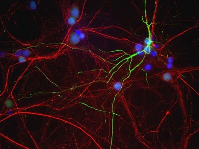
- Mixed neuron/glia cultures from newborn rat brain stained with MCS-7C5 antibody to peripherin (green) and chicken polyclonal antibody to phosphorylated NF-H CPCA-NF-H (red channel). A class of large neurons, like the one in the middle of this image, contain peripherin, while the majority of neurons and their processes contain NF-L and not peripherin. Interestingly, the peripherin positive cells often contain a cytoplasmic inclusion next to the nucleus which stains for both peripherin and NF-L, and so appears golden in this kind of image. The blue channel reveals the localization of DNA.
- Submitted by
- LSBio (provider)
- Main image

- Experimental details
- Mixed neuron/glia cultures from newborn rat brain stained with MCS-7C5 antibody to peripherin (green) and chicken polyclonal antibody to phosphorylated NF-H CPCA-NF-H (red channel). A class of large neurons, like the one in the middle of this image, contain peripherin, while the majority of neurons and their processes contain NF-L and not peripherin. Interestingly, the peripherin positive cells often contain a cytoplasmic inclusion next to the nucleus which stains for both peripherin and NF-L, and so appears golden in this kind of image. The blue channel reveals the localization of DNA.
