Explore
 Validate
 Learn
Learn Western blot
 Flow cytometry
Antibody data
- Antibody Data
- Antigen structure
- References [6]
- Comments [0]
- Validations
- Western blot [1]
- Immunocytochemistry [4]
- Immunohistochemistry [7]
- Other assay [5]
Submit
Validation data
Reference
Comment
Report error
- Product number
- MA5-31980 - Provider product page

- Provider
- Invitrogen Antibodies
- Product name
- CD9 Recombinant Rabbit Monoclonal Antibody (SA35-08)
- Antibody type
- Monoclonal
- Antigen
- Synthetic peptide
- Description
- Recombinant rabbit monoclonal antibodies are produced using in vitro expression systems. The expression systems are developed by cloning in the specific antibody DNA sequences from immunoreactive rabbits. Then, individual clones are screened to select the best candidates for production. The advantages of using recombinant rabbit monoclonal antibodies include: better specificity and sensitivity, lot-to-lot consistency, animal origin-free formulations, and broader immunoreactivity to diverse targets due to larger rabbit immune repertoire.
- Reactivity
- Human, Mouse, Rat
- Host
- Rabbit
- Isotype
- IgG
- Antibody clone number
- SA35-08
- Vial size
- 100 μL
- Concentration
- 1 mg/mL
- Storage
- Store at 4°C short term. For long term storage, store at -20°C, avoiding freeze/thaw cycles.
Submitted references Extracellular Vesicle-Encapsulated MicroRNA-375 from Bone Marrow-Derived Mesenchymal Stem Cells Inhibits Hepatocellular Carcinoma Progression through Regulating HOXB3-Mediated Wnt/β-Catenin Pathway.
Characterization of plasma exosomal microRNAs in responding to radiotherapy of human esophageal squamous cell carcinoma.
Ageing related thyroid deficiency increases brain-targeted transport of liver-derived ApoE4-laden exosomes leading to cognitive impairment.
Genetic Background and Kinetics Define Wound Bed Extracellular Vesicles in a Mouse Model of Cutaneous Injury.
Macrophage secretion of miR-106b-5p causes renin-dependent hypertension.
Inhalation of lung spheroid cell secretome and exosomes promotes lung repair in pulmonary fibrosis.
Yu Z, Liu J, Fan Q, Yu J, Ren X, Wang X
Analytical cellular pathology (Amsterdam) 2022;2022:9302496
Analytical cellular pathology (Amsterdam) 2022;2022:9302496
Characterization of plasma exosomal microRNAs in responding to radiotherapy of human esophageal squamous cell carcinoma.
Miao N, Cai W, Ding S, Liu Y, Chen W, Sun T
Molecular medicine reports 2022 Sep;26(3)
Molecular medicine reports 2022 Sep;26(3)
Ageing related thyroid deficiency increases brain-targeted transport of liver-derived ApoE4-laden exosomes leading to cognitive impairment.
Zhang M, Gong W, Zhang D, Ji M, Chen B, Chen B, Li X, Zhou Y, Dong C, Wen G, Zhan X, Wu X, Cui L, Feng Y, Wang S, Yuan H, Xu E, Xia M, Verkhratsky A, Li B
Cell death & disease 2022 Apr 25;13(4):406
Cell death & disease 2022 Apr 25;13(4):406
Genetic Background and Kinetics Define Wound Bed Extracellular Vesicles in a Mouse Model of Cutaneous Injury.
Qian J, Park DJ, Perrott S, Patel P, Eliceiri BP
International journal of molecular sciences 2021 Mar 29;22(7)
International journal of molecular sciences 2021 Mar 29;22(7)
Macrophage secretion of miR-106b-5p causes renin-dependent hypertension.
Oh J, Matkovich SJ, Riek AE, Bindom SM, Shao JS, Head RD, Barve RA, Sands MS, Carmeliet G, Osei-Owusu P, Knutsen RH, Zhang H, Blumer KJ, Nichols CG, Mecham RP, Baldán Á, Benitez BA, Sequeira-Lopez ML, Gomez RA, Bernal-Mizrachi C
Nature communications 2020 Sep 23;11(1):4798
Nature communications 2020 Sep 23;11(1):4798
Inhalation of lung spheroid cell secretome and exosomes promotes lung repair in pulmonary fibrosis.
Dinh PC, Paudel D, Brochu H, Popowski KD, Gracieux MC, Cores J, Huang K, Hensley MT, Harrell E, Vandergriff AC, George AK, Barrio RT, Hu S, Allen TA, Blackburn K, Caranasos TG, Peng X, Schnabel LV, Adler KB, Lobo LJ, Goshe MB, Cheng K
Nature communications 2020 Feb 28;11(1):1064
Nature communications 2020 Feb 28;11(1):1064
No comments: Submit comment
Supportive validation
- Submitted by
- Invitrogen Antibodies (provider)
- Main image
- Experimental details
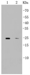
- Western blot analysis of CD9 in different lysates using a Monoclonal antibody (Product #MA5-31980) at a dilution of 1:500. Positive control: Lane 1: Mouse heart, Lane 2: Jurkat.

Supportive validation
- Submitted by
- Invitrogen Antibodies (provider)
- Main image

- Experimental details
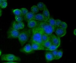
- Immunocytochemical analysis of CD9 in SW480 cells using a CD9 Monoclonal antibody (Product # MA5-31980) as seen in green. The nuclear counter stain is DAPI (blue). Cells were fixed in paraformaldehyde, permeabilised with 0.25% Triton X100/PBS.

- Submitted by
- Invitrogen Antibodies (provider)
- Main image

- Experimental details
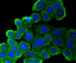
- Immunocytochemical analysis of CD9 in CRC cells using a CD9 Monoclonal antibody (Product # MA5-31980) as seen in green. The nuclear counter stain is DAPI (blue). Cells were fixed in paraformaldehyde, permeabilised with 0.25% Triton X100/PBS.

- Submitted by
- Invitrogen Antibodies (provider)
- Main image
- Experimental details
- Immunocytochemical analysis of CD9 in SW480 cells using a CD9 Monoclonal antibody (Product # MA5-31980) as seen in green. The nuclear counter stain is DAPI (blue). Cells were fixed in paraformaldehyde, permeabilised with 0.25% Triton X100/PBS.

- Submitted by
- Invitrogen Antibodies (provider)
- Main image
- Experimental details
- Immunocytochemical analysis of CD9 in CRC cells using a CD9 Monoclonal antibody (Product # MA5-31980) as seen in green. The nuclear counter stain is DAPI (blue). Cells were fixed in paraformaldehyde, permeabilised with 0.25% Triton X100/PBS.

Supportive validation
- Submitted by
- Invitrogen Antibodies (provider)
- Main image
- Experimental details
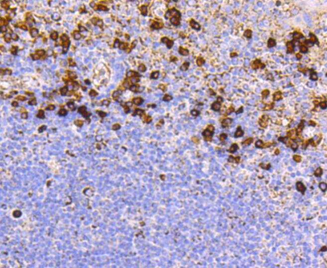
- Immunohistochemical analysis of CD9 of paraffin-embedded Human tonsil tissue using a CD9 Monoclonal antibody (Product #MA5-31980). Counter stained with hematoxylin.
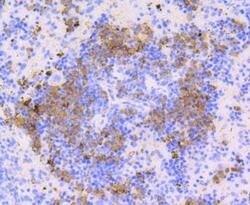
- Submitted by
- Invitrogen Antibodies (provider)
- Main image
- Experimental details
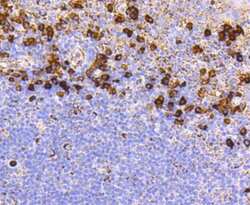
- Immunohistochemical analysis of CD9 of paraffin-embedded Human spleen tissue using a CD9 Monoclonal antibody (Product #MA5-31980). Counter stained with hematoxylin.

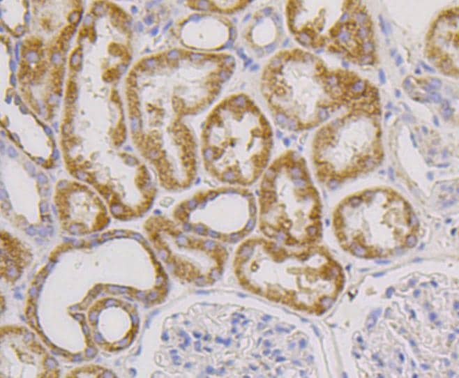
- Submitted by
- Invitrogen Antibodies (provider)
- Main image
- Experimental details
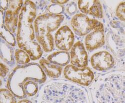
- Immunohistochemical analysis of CD9 of paraffin-embedded Human kidney tissue using a CD9 Monoclonal antibody (Product #MA5-31980). Counter stained with hematoxylin.

- Submitted by
- Invitrogen Antibodies (provider)
- Main image
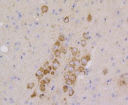
- Experimental details
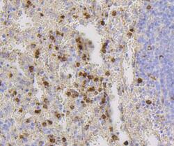
- Immunohistochemical analysis of CD9 of paraffin-embedded Mouse brain tissue using a CD9 Monoclonal antibody (Product #MA5-31980). Counter stained with hematoxylin.

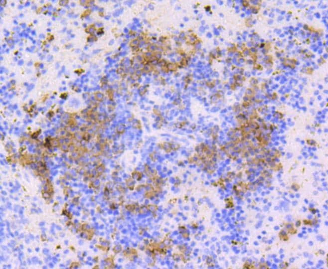
- Submitted by
- Invitrogen Antibodies (provider)
- Main image
- Experimental details
- Immunohistochemical analysis of CD9 of paraffin-embedded Mouse spleen tissue using a CD9 Monoclonal antibody (Product #MA5-31980). Counter stained with hematoxylin.

- Submitted by
- Invitrogen Antibodies (provider)
- Main image

- Experimental details
- Immunohistochemical analysis of CD9 of paraffin-embedded Human kidney tissue using a CD9 Monoclonal antibody (Product #MA5-31980). Counter stained with hematoxylin.
- Submitted by
- Invitrogen Antibodies (provider)
- Main image

- Experimental details
- Immunohistochemical analysis of CD9 of paraffin-embedded Mouse spleen tissue using a CD9 Monoclonal antibody (Product #MA5-31980). Counter stained with hematoxylin.
Supportive validation
- Submitted by
- Invitrogen Antibodies (provider)
- Main image
- Experimental details
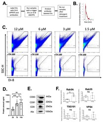
- Figure 5 Characterization of EVs released in the PVA sponge model in C57 BL/6 mice. ( A ) Summary of vFC analysis. ( B ) NTA analysis of size distribution of EVs following ultra-centrifugation. ( C ) Analysis of EVs labeled with di-8 ANEPPS (top row), and gating on Triton-X100 detergent sensitive EVs (bottom row). ( D ) Quantification of protein concentration at 2, 7 and 14 days post-implantation used to normalize vFC analysis of EVs. Data shown are mean +- SD ( n = 5; * p < 0.05, ** p < 0.01, and *** p < 0.001). ( E ) Immunoblotting of EVs to detect Alix, CD9, CD63, and CD81 at 14 days post-implantation of PVA sponges. ( F ) The gene expression analysis of genes associated with EV biogenesis in PVA cells recruited to PVA sponge at 2 and 14 days (* p < 0.05, ns = not significant).
- Submitted by
- Invitrogen Antibodies (provider)
- Main image
- Experimental details
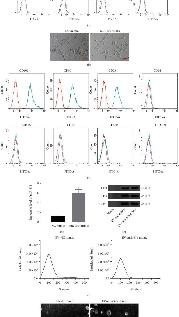
- Figure 2 EV from miR-375 overexpression-modified BM-MSCs are successfully extracted. (a) Expressions of BM-MSC markers CD73, CD90, and CD105 and hematopoietic and endothelial cell markers CD34, CD11b, CD19, CD45, and HLA-DR were measured by flow cytometry. (b). Morphological observation of BM-MSCs. (c) Expression of BM-MSC markers CD73, CD90, and CD105 and hematopoietic and endothelial cell markers CD34, CD11b, CD19, CD45, and HLA-DR in BM-MSCs transfected with NC mimic and miR-375 mimic were measured by flow cytometry. (d) miR-375 expression in BM-MSCs after transfection using RT-qPCR. (e) Expression of EV markers CD9, CD63, and CD81 was determined by Western blot assay. (f) The size distribution of EV analyzed using NTA. (g) TEM observation of EV structure. * p < 0.05 vs. the NC mimic group. All data were expressed as mean +- standard deviation of at least three independent experiments. Data analysis between the two groups was performed by unpaired t -test.
- Submitted by
- Invitrogen Antibodies (provider)
- Main image
- Experimental details
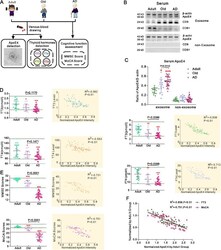
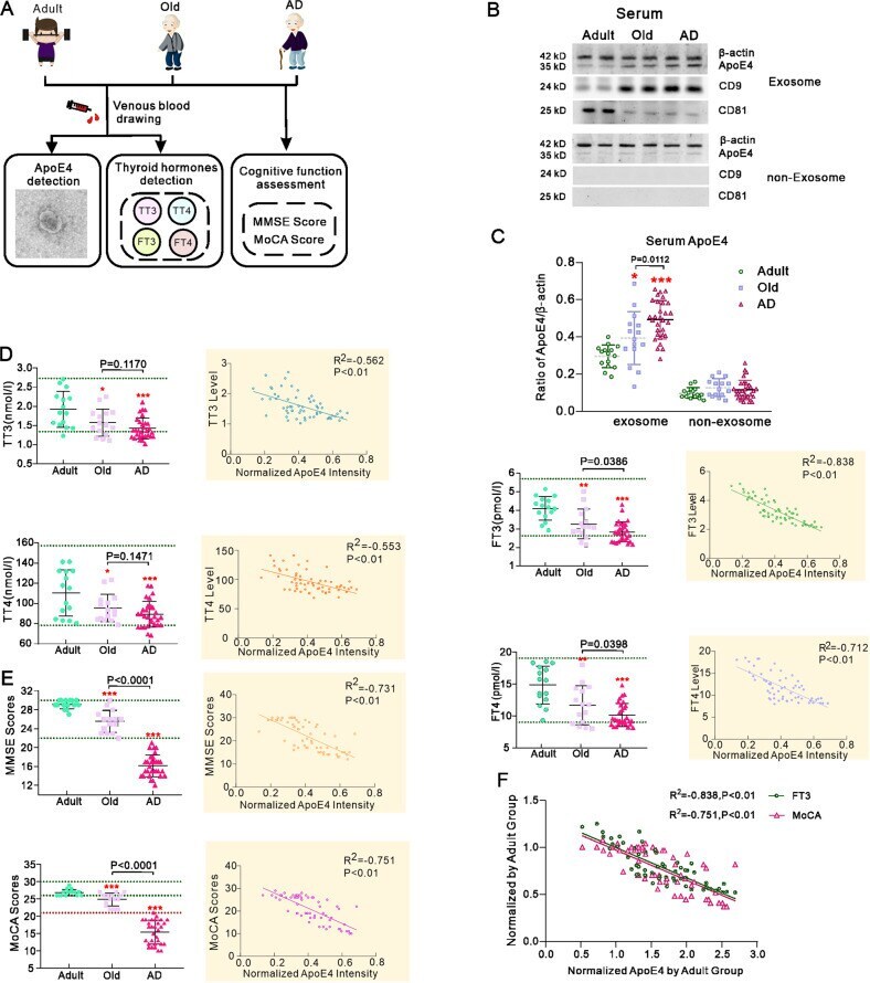
- Correlation of ApoE4 level in serum exosomes with thyroid hormones levels and cognitive performance in human subjects. A Enroled 60 carriers of ApoE4 allele included 15 adult healthy subjects, 15 old healthy subjects and 30 old AD patients. Collected serum was used to extract exosomes and measure TT3, FT3, TT4 and FT4. Participants cognitive abilities were assessed by MMSE and MoCA scoring. B Representative western blots of ApoE4 and exosomal specific marker CD81 and CD9 in the serum extracted exosomes and the supernatant from which exosomes precipitate was removed after ultracentrifuge. C Normalised intensities of ApoE4 by beta-actin. D TT3, FT3, TT4 and FT4 in adult, old and AD groups. Correlation analyses between the ApoE4 level in serum exosomes and TH levels are shown in inserts at the right. E MMSE and MoCA scores; correlation analyses between the ApoE4 level in serum exosomes and these scores are shown in inserts at the right. F Multiple correlation analysis of the normalised ApoE4 in serum exosomes with the normalised MoCA scores and serum FT3 in adult group. The individual measured value was plotted and shown as mean +- SD. One-way ANOVA for comparisons including more than two groups; unpaired two-tailed t -test for two-group comparisons. * p < 0.05, ** p < 0.01, *** p < 0.001.
- Submitted by
- Invitrogen Antibodies (provider)
- Main image
- Experimental details
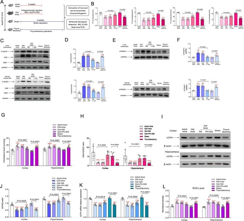
- The effects of L-thy and ApoE4 inhibitor on the exosome transport of h-ApoE4 and cerebral oxidative stress in ApoE4-KI mice. A Old mice were randomly intraperitoneally injected with normal saline (NS), 20 mug/kg/day L-thy or 200 mg/kg/day ApoE4 inhibitor PH-002 for 14 days; 2 months old mice randomly underwent thyroidectomy or sham surgery. Four weeks after surgery measurements were made. B TT3, FT3, TT4 and FT4 values are plotted as mean +- SD, n = 6. One-way ANOVA for comparisons including more than two groups; unpaired two-tailed t -test for two-group comparisons. As comparison with adult group, * p < 0.05, ** p < 0.01, *** p < 0.001. C Representative Western blots of h-ApoE4 in the exosomes extracted from liver and serum, CD9 and CD81 were used as the specific markers of exosomes. D Expression ratio of h-ApoE4 and GAPDH are plotted as mean +- SD, n = 6. E Representative Western blots of ATP6AP1 in the exosomes extracted from liver and serum. F Ratio of ATP6AP1 normalised to GAPDH is plotted as mean +- SD, n = 6. One-way ANOVA for comparisons including more than two groups; unpaired two-tailed t -test for two-group comparisons. As comparison with adult group, * p < 0.05, ** p < 0.01, *** p < 0.001. G , H Cholesterol and the ratio of GSH/GSSG in cortex and hippocampus measured by ELISA are plotted as mean +- SD, n = 6. I Representative Western blots of UCP4 in cortex and in hippocampus. J Ratio of UCP4 to beta-actin is plotted as mean +- SD, n = 6. K mRNA expression of UCP4
- Submitted by
- Invitrogen Antibodies (provider)
- Main image
- Experimental details
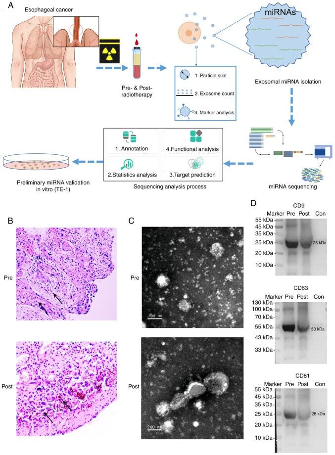
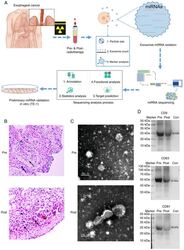
- Figure 1. Exosome miRNA validation and sequencing. (A) The experimental workflow of characterization of plasma exosome miRNAs for the radiotherapy of human esophageal cancer. (B) H&E staining of esophageal squamous cell carcinoma samples (magnification, x100) from patients of pre- and post-radiotherapy. (C) Characterization of plasma exosomes. Morphological observation of exosomes by transmission electron microscopy. Scale bar=200 nm. (D) Expression levels of CD9, CD63 and CD81 were identified using western blot analysis. miRNA, microRNA.