Explore
 Validate
 Learn
Learn Western blot
Antibody data
- Antibody Data
- Antigen structure
- References [2]
- Comments [0]
- Validations
- Western blot [1]
- Immunocytochemistry [2]
- Immunohistochemistry [1]
- Flow cytometry [1]
- Other assay [1]
Submit
Validation data
Reference
Comment
Report error
- Product number
- PA5-11559 - Provider product page

- Provider
- Invitrogen Antibodies
- Product name
- CD9 Polyclonal Antibody
- Antibody type
- Polyclonal
- Antigen
- Synthetic peptide
- Reactivity
- Human, Rat
- Host
- Rabbit
- Isotype
- IgG
- Vial size
- 200 μL
- Concentration
- 0.5 mg/mL
- Storage
- Store at 4°C short term. For long term storage, store at -20°C, avoiding freeze/thaw cycles.
Submitted references Functional Characterization of Human Induced Pluripotent Stem Cell-Derived Endothelial Cells.
Combined Exosomal GPC1, CD82, and Serum CA19-9 as Multiplex Targets: A Specific, Sensitive, and Reproducible Detection Panel for the Diagnosis of Pancreatic Cancer.
Fan X, Cyganek L, Nitschke K, Uhlig S, Nuhn P, Bieback K, Duerschmied D, El-Battrawy I, Zhou X, Akin I
International journal of molecular sciences 2022 Jul 31;23(15)
International journal of molecular sciences 2022 Jul 31;23(15)
Combined Exosomal GPC1, CD82, and Serum CA19-9 as Multiplex Targets: A Specific, Sensitive, and Reproducible Detection Panel for the Diagnosis of Pancreatic Cancer.
Xiao D, Dong Z, Zhen L, Xia G, Huang X, Wang T, Guo H, Yang B, Xu C, Wu W, Zhao X, Xu H
Molecular cancer research : MCR 2020 Feb;18(2):300-310
Molecular cancer research : MCR 2020 Feb;18(2):300-310
No comments: Submit comment
Supportive validation
- Submitted by
- Invitrogen Antibodies (provider)
- Main image

- Experimental details
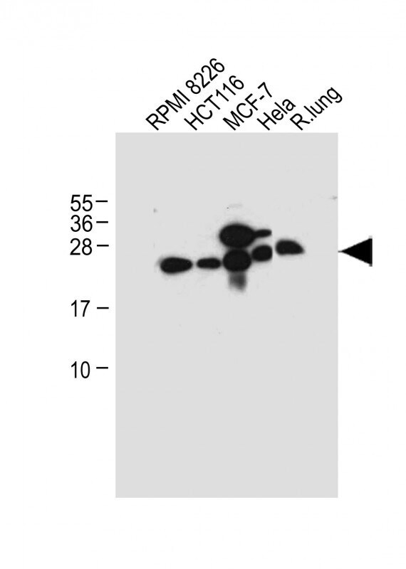
- Western blot analysis of CD9 in various lysates. Samples were incubated with CD9 polyclonal antibody (Product # PA5-11559) using a dilution of 1:1,000 followed by Goat Anti-Rabbit IgG, (H+L), Peroxidase conjugated at a dilution of 1:10,000. Lysates/proteins: 20 µg per lane. Lane 1: RPMI 8226 whole cell lysate; Lane 2: HCT116 whole cell lysate; Lane 3: MCF-7 whole cell lysate; Lane 4: Hela whole cell lysate; Lane 5: Rat lung tissue lysate. Predicted band size: 25 kDa. Blocking/Dilution buffer: 5% NFDM/TBST.
Supportive validation
- Submitted by
- Invitrogen Antibodies (provider)
- Main image
- Experimental details
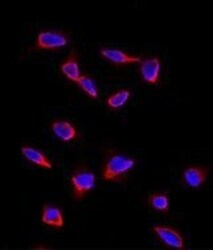
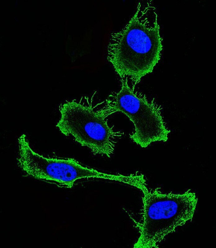
- Immunofluorescent analysis using a CD9 polyclonal antibody (Product # PA5-11559) in HeLa cells. 0.025 mg/mL primary antibody was followed by fluor-conjugated donkey anti-rabbit lgG (H+L) (orange). Blue counterstaining is DAPI.

- Submitted by
- Invitrogen Antibodies (provider)
- Main image
- Experimental details
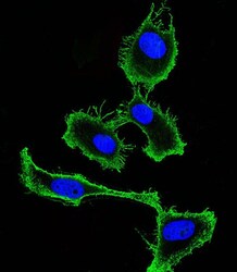
- Immunocytochemistry analysis of CD9 in Hela (Human Cervical epithelial adenocarcinoma cell line) cells. Samples were incubated with CD9 polyclonal antibody (Product # PA5-11559) using a dilution of 1:25 followed by Dylight® 488-conjugated goat anti-rabbit IgG at a dilution of 1:200 (green). Cells were 4% paraformaldehyde-fixed and 0.1% Triton X-100 permeabilized. Immunofluorescence image showing membrane staining on Hela cell line. The nuclear counter stain is DAPI (blue).
Supportive validation
- Submitted by
- Invitrogen Antibodies (provider)
- Main image
- Experimental details
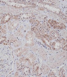
- Immunohistochemistry analysis of CD9 in paraffin-embedded Human kidney tissue. Samples were incubated with CD9 polyclonal antibody (Product # PA5-11559) using a dilution of 1:100 for 1 hour at room temperature followed by undiluted CRF Anti-Polyvalent HRP Polymer antibody. Tissue was fixed with formaldehyde at room temperature. Heat induced epitope retrieval was performed by EDTA buffer (pH 9 0).

Supportive validation
- Submitted by
- Invitrogen Antibodies (provider)
- Main image
- Experimental details
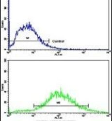
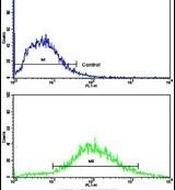
- Flow cytometry analysis of Jurkat cells using a CD9 polyclonal antibody (Product # PA5-11559) (bottom), compared to a negative control cell (top) at a dilution of 1:10-50, followed by a FITC-conjugated goat anti-rabbit antibody
Supportive validation
- Submitted by
- Invitrogen Antibodies (provider)
- Main image
- Experimental details
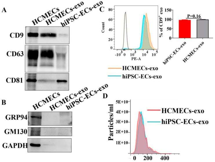
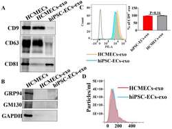
- Characterization of the purified exosomes from hiPSC-EC and HCMEC conditioned media. Exosomes isolated from cell culture media of hiPSC-ECs and HCMECs. ( A ) Western blot showed the detection of exosome markers CD9, CD63, and CD81 in isolated exosomes. ( B ) Glucose-regulated protein 94 (GRP94) and Golgi marker GM130 were not found in exosomes. ( C ) Flow cytometry analysis using exosome marker CD9. ( D ) The size distribution of isolated exosomes was measured by nanoparticle tracking analysis (NTA) in hiPSC-ECs and HCMECs.